Lower Limb
Nerves, Blood Vessels and Lymph
The Thigh
Nervous Supply
The sciatic nerve is the largest single nerve in the human body; it runs from each side of the lower spine through deep in the buttock into the back of the thigh and all the way down to the foot. It serves a vital role in connecting the spinal cord with the leg and foot muscles. The sciatic nerve is derived from the lumbosacral plexus. After its formation, it leaves the pelvis and enters the gluteal region via greater sciatic foramen. It emerges inferiorly to the piriformis muscle and descends in an inferolateral direction. As the nerve moves through the gluteal region, it crosses the posterior surface of the superior gemellus, obturator internus, inferior gemellus and quadratus femoris muscles. It then enters the posterior thigh by passing deep to the long head of the biceps femoris. Within the posterior thigh, the nerve gives rise to branches to the hamstring muscles and adductor magnus. When the sciatic nerve reaches the apex of the popliteal fossa, it terminates by bifurcating into the tibial and common fibular nerves. Any type of pain and/or neurological symptoms that are felt along the sciatic nerve is referred to as sciatica.
The saphenous nerve is a sensory branch of the femoral nerve and supplies sensation to the anteromedial, medial and posteromedial surface of the leg. The nerve passes through the adductor canal and gives off an infrapatellar branch. It continues to become subcutaneous to supply prepatellar skin and also supplies the medial side of the ankle and foot.
The obturator nerve is formed by the anterior divisions of the second, third and fourth lumbar nerves. It descends through the fibers of the psoas major muscle and emerges from its medial border, running posteriorly to the common iliac arteries and laterally along the pelvic wall to the obturator foramen. It then enters the thigh through the obturator canal and splits into anterior and posterior divisions. The anterior division descends between the adductor longus and adductor brevis muscles towards the femoral artery, giving off branches to the adductor longus, adductor brevis and gracilis muscles. In rare cases, it also gives off a branch to the pectineus muscle which then pierces the fascia lata to become the cutaneous branch of the obturator nerve. The posterior division descends through the obturator externus muscle before passing anteriorly to adductor magnus and giving off branches to supply it.
Arterial Supply
The perforating artery originates in the deep part of the thigh and distributes to three or four vessels that pass through the great adductor muscle to the posterior and lateral parts of the thigh.
The descending genicular artery is found in the anterior portion of the thigh. It branches off from the femoral artery and then immediately splits into the saphenous branch and the articular branches of the descending genicular artery. The descending genicular artery rarely has an aneurysm, which is a ballooning of weak blood vessel walls. In some cases, this artery is used as a bypass route when other blood vessels of the leg develop blood clots or other blockages.
Venous Supply
The great saphenous vein is a large venous blood vessel running near the inside surface of the leg from the ankle to the groin. It arises from the dorsal venous arch at the top of the foot and drains into the femoral vein, the main deep vein for the leg. The great saphenous vein is sometimes stripped out of the leg to eliminate varicose veins. It is also used as a source of grafts in coronary bypass surgery.
Perforator veins are small veins that connect the deep and superficial systems together. They are usually short in length and have only a few valves in them. They can have reflux just like other veins in the legs. These small connector veins run horizontally across the leg and not vertically up and down the leg. A visual representation would be compared to a horizontal cross on the letter H. These perforators can be the source to visible varicose veins in the calf or thigh.
Lymph Supply
The lymphatic system functions to drain tissue fluid, plasma proteins and other cellular debris back into the blood stream, and is also involved in immune defence. Once this collection of substances enters the lymphatic vessels it is known as lymph; lymph is subsequently filtered by lymph nodes and directed into the venous system.
The superficial vessels can be divided into two major subsets; (i) medial vessels, which closely follow the course of the great saphenous vein and; (ii) lateral vessels which are more closely associated with the small saphenous vein.
Medial Vessels: The medial group originate on the dorsal surface of the foot. They travel up the anterior and posterior aspects of the medial lower leg, with the great saphenous vein, passing with it behind the medial condyle of the femur. This group of vessels ends in the groin, draining into the sub inguinal group of the inguinal lymph nodes.
Lateral Vessels: The lateral vessels arise from the lateral surface of the foot and either accompany the small saphenous vein to enter the popliteal nodes, or ascend in front of the leg and cross just below the knee joint to join the medial group.
The Knee
Nervous Supply:
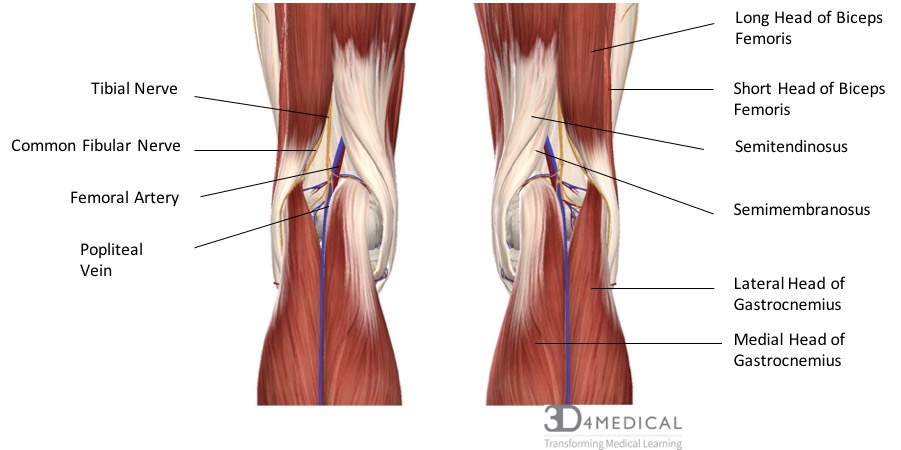
Traveling through the popliteal region include some of the major nerves, arteries, veins, and lymph that continue down the posterior side of the leg. The two major nerves include the tibial nerve and the common fibular nerve. The tibial nerve arises from the sciatic nerve which splits just superior to the knee. The tibial nerve splits into an articular branch which works along with the obturator nerve for sensation to the knee, and a muscular branch which supplies all the muscles in the posterior compartment of the leg including gastrocnemius, soleus, plantaris, popliteus, tibialis posterior, flexor digitorum longus, and flexor halluces longus. The common fibular nerve, also called the peroneal nerve, arises from the lateral aspect of the sciatic nerve. It runs along the lateral condyle of the femur and then passes under fibularis longus where it then branches into the superficial fibular nerve and the deep fibular nerve. The common fibular nerve supplies the short head of biceps femoris in the thigh and its terminal branches supply all the muscles in the anterior and lateral compartments of the leg.
Arterial Supply:
The main artery traveling through the popliteal fossa is the femoral artery which turns into the popliteal artery when it passes over the tibiofemoral joint. The femoral artery passes through the adductor hiatus in adductor magnus before entering the popliteal fossa and turning into the popliteal artery. The popliteal artery is the primary distributor of oxygenated blood to regions around the knee. Within the popliteal fossa the popliteal artery branches into other significant blood vessels such as the sural artery and the various types of genicular arteries. At the distal end, the popliteal artery splits into the anterior and posterior tibial arteries which supply oxygenated blood to the lower leg.
Venous Supply:
Running parallel to the popliteal artery is the popliteal vein which is formed from the anterior and posterior tibial veins. The popliteal vein is responsible for returning blood from the calf, the knee, and the thigh back to the heart. The small saphenous vein which originates in the foot empties into the popliteal vein as well as many of the genicular veins and the sural vein which surround the knee.
Lymphatic Supply:
Within the popliteal fossa, there are many different lymph nodes and ducts. The main nodes in this region include the deep and superficial popliteal lymph nodes which are supplied by the medial superficial lymph vessels of the leg. These superficial lymph vessels lie within the superficial fascia of the leg and can be divided into a lateral and medial group. Both the deep and superficial lymph nodes and their vessels work together to drain the lower leg of lymph.
Nerves of the Foot
There are many nerves that work to give sensation and reception to the feet. These include the tibial nerve, the common peroneal nerve, sural nerve, saphenous nerve, medial plantar nerve, lateral plantar nerve, plantar digital nerve and the calcaneal branches of the tibial and sural nerve. They are all used for the innervation of different parts and sections of the foot.
The tibial nerve runs posteriorly in the leg through the two heads of the gastrocnemius and loops underneath the medial malleolus to continue into the bottom of the foot. The purpose of the tibial nerve is to innervate all of the muscles in the posterior compartment of the leg. The common peroneal nerve is unique in the fact that it branches off into two nerves; the superficial fibular nerve and the deep fibular nerve. The superficial peroneal nerve innervates the muscles in the lateral compartment of the leg and gives sensation to part of the lateral ankle. The deep peroneal nerve runs down beside flexor digitorum longus and innervates the muscles in the anterior compartment of the leg. The sural nerve runs down to the medial part of the foot, innervating the skin on the lateral side of the leg and foot and is formed by branches from the tibial nerve and the common peroneal nerve. The saphenous nerve is a branch off of the femoral nerve and it innervates the skin medial side of the foot. The medial plantar nerve branches off of the tibial nerve and innervates some of the medial muscles in the foot, as well as the skin of the medial plantar region of the foot. The lateral plantar nerve is also a branch off of the tibial nerve and innervates several small muscles in the foot as well as innervating the skin on the lateral side of the plantar region. Both the medial and lateral plantar nerves have branches going off of them to form the plantar digital nerve. The plantar digital nerve innervates the skin of the toes. The calcaneal branches of the tibial and sural nerve location is named after its name, where it runs down the back of the leg to innervate the heel skin of the heel.
