Introduction
Surface Anatomy
LATERAL CERVICAL REGION
The lateral cervical region has three main borders: the mandible, the internal jugular vein, and the clavicle. As shown in the figure above, the region is inferior to the mandible, anterior to the internal jugular vein, and superior to the clavicle. This region includes lots of muscles, nerves, arteries and veins, and lymph. It is also important to note that this region is essential to brain function as it has a large amount of blood supply as well as nerves. For those reasons, this area is very vulnerable.

SURFACE ANATOMY OF THE LATERAL CERVICAL REGION
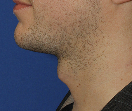
The surface of the anterior cervical region is commonly referred to as the throat. The throat has one very distinctive feature that is commonly called the Adam’s apple. This feature is created by the tissue size of the larynx. Women tend to have smaller larynx’ which is why the Adam’s apple is much more prominent in men.
POSTERIOR CERVICAL REGION
For the posterior cervical region, important surface anatomy used in a clinical setting include the spinous processes of the cervical vertebrae (follow link to bones of the head and neck). C1 has no spinous process, so the first palpable landmark on the cervical spine is the is C2. While, inferiorly along the cervical spine, C7 is distinct with non-bifid spinous process and is rather bulbous, creating a visible bump on the surface. C7 is called the vertebral prominens. The position transverse processes are used to map out where to find muscles and nerves in the region. Other bony landmarks include:
- External Occipital Protuberance which lies in the middle and the base of the occipital bone and which are important attachment sites for the trapezius and the nuchal ligament.
- Nuchal lines (Superior and Inferior) are four ridges that flank either side of the occipital protuberance and run horizontally toward the lateral portion of the skull. They are important sites for muscles, such as splenius capitis, semispinalis capitis, longissimus capitis and the trapezius, attaching the skull to the vertebral column. The occipital belly of the occipitofrontalis muscle attaches to the superior nuchal lines. The nuchal lines are important clinically as an indicator area for pain.
Posterior Cervical Triangle is an area of vulnerability due to important nerves, arteries and veins travelling through the neck to reach the head and are present near the surface of the body making them exposed to weakness.
The posterior cervical triangle has its superior angle between the mastoid process and occipital protuberance and is bordered by the sternocleidomastoid anteriorly, the upper trapezius posteriorly, and the clavicle forms the inferior border. Within the triangle are portions of the splenius capitis, levator scapulae, middle scalene, and the posterior scalene that wraps around to the lateral surface. The omohyoid crosses the posterior triangle. The floor is formed by deep cervical fascia. 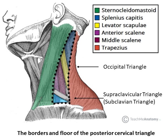
The posterior cervical triangle can be divided into the large upper Occipital Triangle, and the small lower Supraclavicular Triangle. The occipital triangle contains a portion of the cranial Accessory Nerve (XI), cervical plexus (posterior branches), brachial plexus (trunks) and is named after the occipital artery which enters the posterior cervical triangle at the superior angle and ascends over the head. The occipital artery is a branch of the external carotid artery. Also a part of the triangle, are the transverse cervical artery and the cervical lymph nodes. The Supraclavicular Triangle is also known as the subclavian triangle because the subclavian artery passes through this area on a deep level. More superficially, this region also includes part of the subclavian vein, suprascapular artery and the external jugular vein. The Supraclavicular Triangle is located in the supraclavicular fossa, the dip above the clavicle from a surface perspective. The Phrenic Nerve (part of the cervical plexus) is an important nerve passing through this region and is often affected by Thoracic Outlet Syndrome.
