Lower Limb
Chapter Conclusion
Learning Objectives
- Identify the femur, tibia, and fibula and their anatomical features.
- Identify the bones of the foot and their relative positions.
- Be able to explain the origin, insertion, action, and innervation of the 5 adductor muscles, the knee flexors and extensors, and muscles that move the foot. (not as much emphasis on muscles that move the foot).
- Identify the seven major supporting ligaments of the knee cap. This includes their function and relative positioning.
- Develop a basic understanding of the ligament in the foot, and ligaments associated with the ankle.
- Identify the major blood vessels in this region and what areas these vessels drain/supply blood to.
- Develop a general understanding of the major clinical conditions in this area.
Part A.
1. The gluteal tuberosity extends from the ___________ & the pectineal line extends from the _____________.
a) greater trochanter, lesser trochanter
b) greater tubercle, lesser tubercle
c) lesser trochanter, greater trochanter
d) lesser tubercle, greater tubercle
2. Which of the following features is not part of the distal epiphysis of the femur?
a) adductor tubercle
b) medial & lateral condyles
c) medial & lateral epicondyles
d) popliteal surface
3. Which of the following bones is the attachment point for the achilles tendon?
a) talus
b) calcaneus
c) navicular
d) cuboid
4. Which of the following adductor muscles inserts on the adductor tubercle?
a) adductor brevus
b) adductor longus
c) adductor magnus
d) all of the above
5. The knee extensor muscles are group together and commonly referred to as the ____________ and insert on the __________ via the patellar ligament.
a) hamstrings, tibial tuberosity
b) hamstring, fibular head
c) quadriceps, fibular head
d) quadriceps, tibial tuberosity
6. Which of the following statements is false when describing the bicep femurs muscle?
a) It’s main actions include knee flexion, hip extension & lateral rotation
b) The long head originates on the ischial tuberosity and the short head originates on the linea aspera
c) both the long and short head insert on the head of the fibula
d) all of the above statements are true
7. How many extrinsic ligaments of the knee joint are there?
a) 2
b) 5
c) 7
d) 9
8. Which of the following plantar flexion muscles is not deep?
a) tibialis posterior
b) flexor digitorum longus
c) flexor hallucis longus
d) all of the above are deep
9. The femoral artery turns into the ____________ at the ______________.
a) femoral artery, popliteal fossa
b) popliteal artery, popliteal fossa
c) anterior tibial nerve, femoral triangle
d) posterior tibial nerve, femoral triangle
10. Osgood-Schlatter disease can best be described as?
a) injury that occurs over the lateral aspect of the knee
b) pain in the front of the knee or around the patella
c) stress on the tibial tuberosity during a growth spurt
d) when a muscle is stretched beyond its limits, tearing its muscle fibers
Part B.
For the following true and false questions, circle T if the answer is true, and F if the answer is false.
- The posterior shaft of the femur contains the lines alba. T / F
- The talus is the largest tarsal bone. T / F
- Sartorious is a unique muscle because it can cause knee and hip flexion. T / F
- All of the quadricep muscles are innervated by the femoral nerve. T / F
- The medial and lateral menisci help prevent medial and lateral displacement of the tibia. T / F
- The muscle in the anterior compartment of the leg cause plantar flexion. T / F
- Gastrocnemius is deep to soleus. T / F
- Adductor brevus, longus, and magnus are all innervated by the Obturator nerve. T / F
- The great and small saphenous veins are deep veins. T / F
- The 3 borders of the femoral triangle include the inguinal ligament, adductor longus, and sartorius. T / F
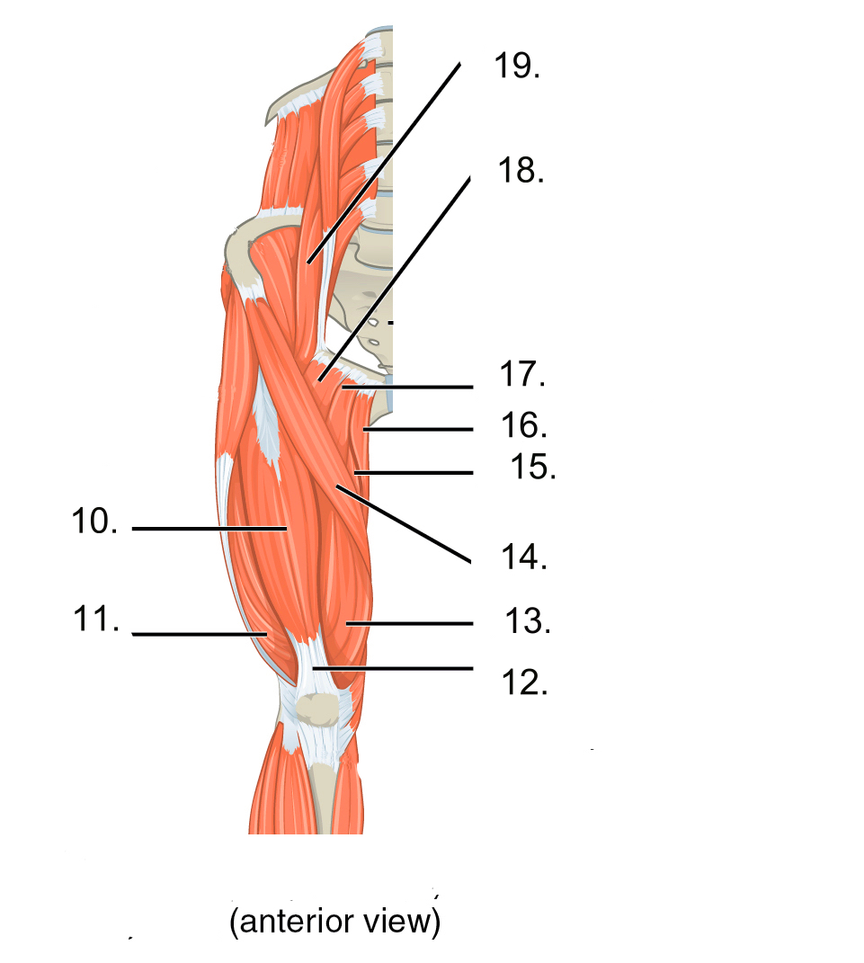
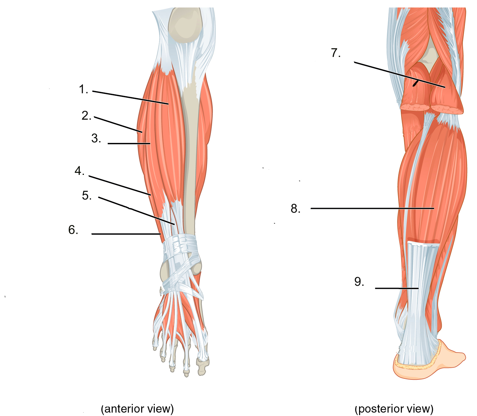
Part C.
Correctly identify the components in the following diagrams. All of the definitions will be used once.
| Questions | Word back |
| 1.
2. 3. 4. 5. 6. 7. 8. 9. 10. 11. 12. 13. 14. 15. 16. 17. 18. 19. |
A. soleus
B. adductor magnus C. pectineus D. rectus femoris E. tibialis anterior F. Psoas major G. gracilis H. gastrocnemius I. quadricep tendon J. calcaneal tendon K. sartorius L. fibularis tertius M. vastus medialis N. vastus lateralis O. adductor longus P. extensor digitorum longus Q. extensor hallucis longus R. fibularis longus S. fibularis brevis |
Answer Key
Part A.
- a 2. c 3. b 4. c 5. a 6. d 7. b 8. d 9. b 10. c
Part B.
- F 2. F 3. T 4. T 5. F 6. F 7. F 8. T 9. F 10. T
Part C.
- E 2. R 3. P 4. S 5. Q 6. L 7. H 8. A 9. J 10. D 11. N 12. I 13. M 14. K 15. B 16. G 17. 0 18. C 19. F
