Upper Limb
The Hand
Articulating Surfaces of the Wrist
The wrist consists of a series of short bones that are located distal to the ulna and radius, and proximal to the metacarpals. Within this small area of bones, there are various articulating surfaces within the wrist. The articulation between the proximal row of carpals and the radius is known as the radiocarpal joint; the articulations in between the carpals are known as the intercarpal joints; and, the articulation between the distal row of carpals and phalanges is known as the carpometacarpal joint.
| Joint | Ligaments | Articular Components | Classification | Movements |
| Radiocarpal | Palmar Radiocarpal Palmar Ulnocarpal Dorsal Radiocarpal Ulnar Collateral Radial Collateral |
Between distal end of: radius and scaphoid, triquetrum, and lunate of carpus. | Structural: synovial Functional: freely moveable |
Slight hyperextension, flexion/extension, abduction/adduction, and circumduction at wrist. |
| Intercarpal | Dorsal and Palmar Interosseous Pisohamate and PisometacarpalCollateral |
Between both rows of carpal bones (midcarpal), and between distal and proximal rows of carpal bones. | Structural: synovial, except for scaphoid, lunate, and hamate which are synovial (saddle). Functional: freely moveable |
Flexion/extension, abduction/adduction, and gliding at midcarpal joints. |
| Carpometacarpal | Dorsal
Palmar Interosseous |
Carpometacarpal joints of remaining digits formed between 2nd-5th metacarpals and carpus; and, carpometacarpal joint of thumb between first metacarpal and trapezium. | Structural: synovial (plane) at digits, synovial (saddle) at thumb. Functional: freely moveable |
Flexion/extension, abduction/adduction, and circumduction at thumb, and gliding at remaining digits. |
Carpal Bones
The carpal bones are the eight bones located within the wrist and form the radiocarpal, intercarpal, and carpometacarpal joints as stated above. The carpal bones allow for limited flexion, extension, radial deviation, and ulnar deviation of the wrist. From an anterior view of the wrist in anatomical position, the eight carpal bones are easily identifiable. The proximal row of four carpal bones listed from lateral to medial are the Scaphoid, Lunate, Triquetrum, and Pisiform. The distal row of carpal bones listed from lateral to medial are the Trapezium, Trapezoid, Capitate, and Hamate. A helpful mnemonic device that lists the bones in this order is: “Sam Likes To Push The Toy Car Hard.”
Metacarpals
Distal to the carpal bones lies the metacarpus (meta = beyond), or palm, the intermediate aspect of the hand; this area is comprised of five bones called metacarpals. Each metacarpal bone is made up of by a proximal base, intermediate shaft, and a distal head – the distal heads are also readily available when one clenches their fists, showing off “knuckles.” Starting with the thumb and moving medially, the bones are numbered I to V. The proximal base articulates with the distal row of carpal bones to form the carpometacarpal joints; and, at the distal heads, they articulate with the phalanges to form the metacarpophalangeal joints.
Phalanges
Distal to the metacarpals are the phalanges (phalanx = a battle line), or rather, the bones of the digits. Again, like the metacarpals they are numbered I to V, moving from lateral to medial. There are a total of fourteen phalanges in the five digits of each hand; and, a single bone of a digit is referred to as a phalanx. Like the metacarpals, the digits are composed of a proximal base, intermediate shaft, and distal head. The thumb has two phalanges, whereas the rest of the digits have three. The phalanges are then broken down into rows, proximal, medial, and distal rows that of which respectively articulate with one another to form interphalangeal joints.
| Joint | Ligaments | Articular Components | Classification | Movements |
| Metacarpophalangeal | Palmar
Deep Transverse Metacarpal |
Between metacarpal heads, and bases of proximal phalanges. | Structural: synovial.
Functional: freely movable. |
Circumduction, flexion/extension, and abduction/adduction. |
| Interphalangeal | Interphalangeal | Between heads of and bases of more distal phalanges. | Structural: synovial (hinge).
Functional: freely movable. |
Flexion/extension of phalanges. |
Extrinsic and Intrinsic Ligaments of the Wrist
Extrinsic wrist ligaments are ligaments that are responsible for attaching and stabilizing the carpal bones to the radius, ulna, and metacarpals. These ligaments can be found on both the dorsal and palmar surfaces of the carpal bones. They have attachments sites on the rough outer layer of the bones called periosteum. The Radioscaphocapitate ligament attaches on the palmar side of the scaphoid and runs laterally, attaching on the styloid process of the radius. The Ulnocapitate ligament attaches on the palmar side of the capitate and runs medially, attaching on the styloid process of the ulna. The Radial collateral ligament attaches on the medial end of the scaphoid and on the styloid process of the radius. The Long Radiolunate ligament attaches on the palmar surface of the lunate and runs laterally, attaching on the middle part of the distal end of the radius. The Short Radiolunate ligament attaches on the lunate and on the medial side of the distal end of the radius. The Ulnolunate ligament attaches on the lunate and runs medially to attach on the ulna. The Ulnotriquetral ligament has attachments on the triquetrum and the distal end of the ulna. A great image to help visualize these ligaments can be found here.
Intrinsic wrist ligament differs from extrinsic ligaments in that they attach carpal bone to carpal bone, anchoring them to each other. The ligaments are named based on which carpal bones they attach to. This makes it very simple to identify ligaments based on their name. Starting laterally on the distal row of carpal bones is the Trapeziotrapezoid ligament. This ligament attaches to the trapezium and trapezoid. Moving medially, the next ligament is the Capitotrapezoid ligament which attached on the Capitate and trapezoid. The next ligament is the Capitohamate ligament attaching to the Capitate and Hamate. The next, more proximal, row of intrinsic ligaments moving from lateral to medial are as follows. The Scaphotrapeziumtrapezoid ligament attaches to the distal end of the Scaphoid and proximal ends of the trapezium and trapezoid. The Scaphocapitate ligament attaches to the distal-medial aspect of the Scaphoid and the proximal-lateral aspect of the Capitate. The Triquetrocapitate attaches to the medial side of the Capitate and the lateral side of the Triquetrum. The Triquetrohamate attaches to the proximal-medial side of the Hamate and the distal end of the Triquetrum. Lastly, the most proximal row of intrinsic carpal ligaments from lateral to medial are the Scapholunate ligament which has attachment points on the medial side of the Scaphoid and lateral side of the Lunate, and the Lunotriquetral ligament which attaches on the medial side of the Lunate and lateral side of the Triquetrum. Below is a great image that helps to understand these ligaments.

Ligaments and Tendons Responsible for Finger Flexion
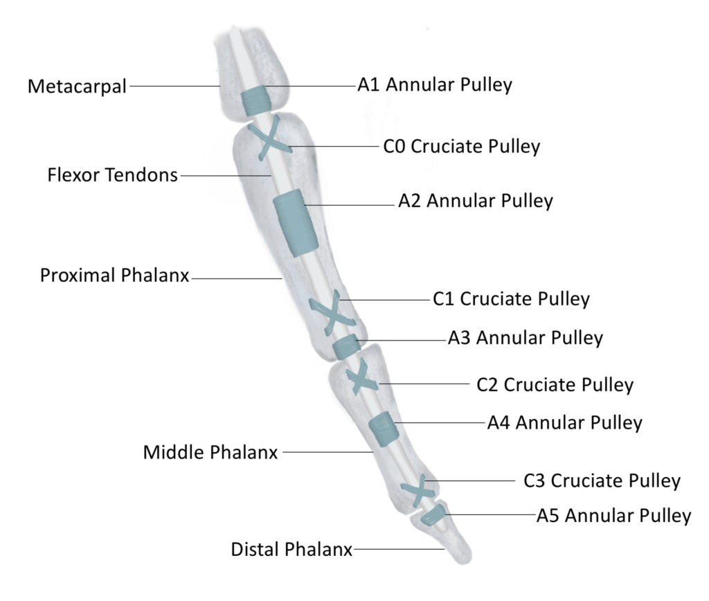
There are two muscles that control the flexion movement of the fingers. This motion would be that of making a fist or closing your hand. These muscles are flexor digitorum Superficialis and Flexor digitorum profundus. Flexor digitorum superficialis originates at the proximal end of the forearm, so on the medial epicondyle, on the radius and ulna. It runs down the forearm, passes deep to the transverse carpal ligament (which is your carpal tunnel) and inserts on the middle phalanx of digits 2-5. Flexor digitorum profundus originates in numerous spots on the medial side of the ulna, and inserts on the base of the distal phalanx of digits 2-5. Flexor digitorum profundus travels through, and past splits of flexor digitorum superficialis. These tendons travel deep to specialized ligaments which create pulleys in the fingers. There are five sets of anular fibers which are referred to as the pulleys. They are called A1-A5. A1 is at the base of the fingers. It is on the metacarpals, right before the proximal phalange. A2 is in the middle of the proximal phalange. A3 is right on the joint between the proximal and middle phalange. A4 is in the middle of the middle phalange, and A5 is on the proximal phalange. The annular pulleys provide primary support to keep the tendons of flexor digitorum superficialis and flexor digitorum profundus close to the bone. Structurally, they provide a ring of connective tissue to hold the tendons in place. Pulleys A2 and A4 are the largest pulleys. They also must endure much higher forces from the tendons. These two pulleys are much more prone to damage than the other smaller pulleys, making them the primary focus of clinical applications.
There are also pulleys called cruciate or cruciform pulleys that provide additional support to the annular pulleys. These are structurally different in that they form an X across the tendon, holding it in place. Each cruciate pulley has four attachment sites on the bone (two on each side) whereas the annular pulleys have two (one on each side). The cruciate pulleys are located in between each of the annular pulleys and are numbered C0 to C3 with C1 being the most proximal and C3 being the most distal. The picture below is of the annular and cruciate pulleys holding the tendons of flexor digitorum profundus and flexor digitorum superficialis close to the bone.
Ligaments and Tendons Responsible for Finger Extension
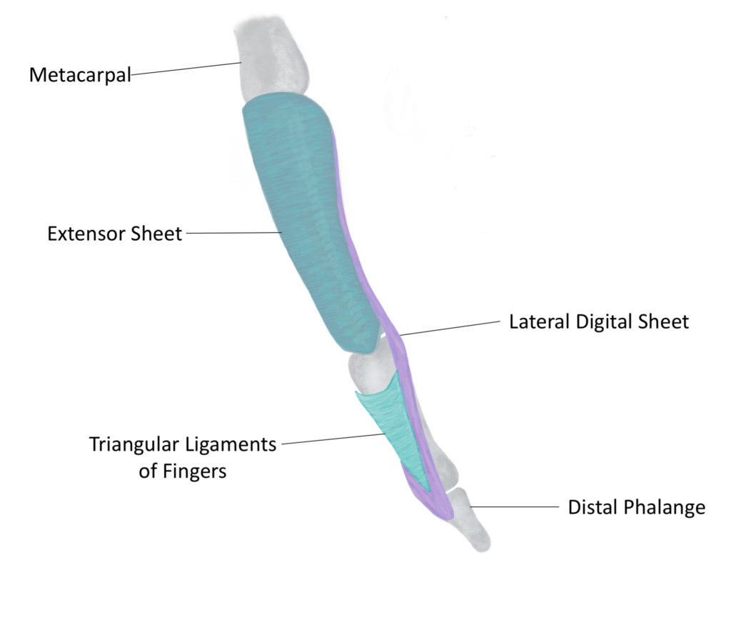
The ligaments of the digits which hold the flexor tendons close to the bone are much more complex than the ligaments which contain the extensor tendons of the digits. This is because the flexor tendons produce large forces against these ligaments whereas the extensor tendons do not. The Annular and Cruciate pulleys are required to keep the flexor tendons close to the bone, opposing their forces. In contrast, the extensor tendons do not pull away from the bone and do not require the same structural support to keep them in place. The tendons that expand to the digits are extensor digitorum, extensor digiti minimi, and extensor indicis. Extensor digiti minimi functions to extend the 5th digit, extensor indicis functions to extend the 2nd digit, and extensor digitorum works to extend and abduct digits 2-5. The three primary ligaments responsible for holding these tendons in place are the extensor sheet, the lateral digital sheet, and the triangular ligaments of the fingers. The extensor sheets span from the base of the distal end of the metacarpal bones to the proximal end of the distal phalanges. They wrap around the lateral sides of the digits to encapsulate the extensor tendons. The lateral digital sheets attach to the perimeter of the extensor sheet and extend to the lateral aspects of each phalange. However, they extend all the way to the distal phalanx and meet on the posterior surface forming a V shape. The triangular ligaments of the fingers span between the two sides of the lateral digital sheets on each phalange. As indicated by the name they form a triangular shape and are located on the posterior side of the digits superficial to the middle phalanx. Below is a photo of the aforementioned ligaments.
Ligaments of the Digits
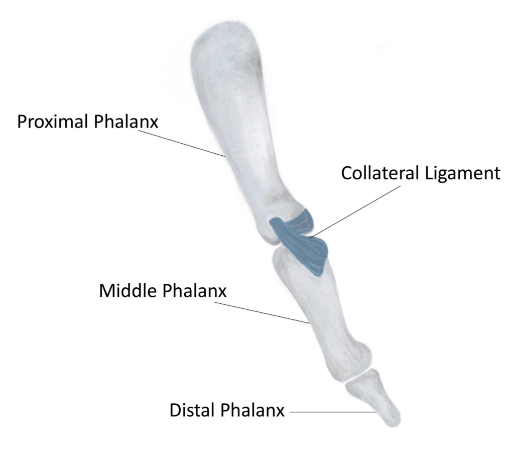
The joints of the digits allow for movement in one plane. The digits can flex and extend, but cannot perform rotation or lateral movement. Each joint has specialized ligaments to prevent abnormal movement to avoid injury. Each joint has two Collateral ligaments associated with it. They are located on the lateral sides of each joint and prevent lateral movement. The PIP joint contains a specialized ligament called the Volar plate. This ligament is on the anterior surface of the digits and connects the proximal and middle phalanx. The Volar plate prevents hyperextension of the PIP joint and provides stability. The picture below represents the PIP collateral ligament from a lateral view.