The Head and Neck
Muscles
The Muscles of the Cranium
| Muscle | Origin | Insertion | Action | Innervation |
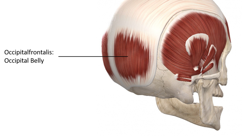
| Occipitalfrontalis | Frontal Belly: Epicranial aponeurosis
Occipital Belly: Superior nuchal line of occipital bone |
Frontal Belly: Skin of the eyebrow and the forehead
Occipital Belly: Epicranial aponeurosis |
Both: elevates the eyebrow, wrinkles the skin of the forehead, and moves the scalp | Frontal Belly: temporal branches of the facial nerve
Occipital Belly: Posterior auricular nerve |
| Temporalis | Temporal fossa; Temporal fascia | Ramus and coronoid process of the mandible | Vertical part: elevation of the mandible
Horizontal part: retraction of the mandible; chewing |
Anterior branch of the deep temporal nerve, posterior branch of deep temporal nerve |
| Temporoparietalis | Fascia superior to the auricular | Anterolateral epicranial aponeurosis | Draws the epicranial aponeurosis towards the front of the cranium |
Temporal branches of the facial nerve |

Epicranial Aponeurosis also referred to as the Galea Aponeurotica or the aponeurosis epicranialis is a broad tendon and is the intermediate section of the occipitofrontalis muscle. It runs from the superior portion of the frontal bone and covers the parietal bones to the lambdoid suture. It is in between the frontalis muscle that lies on the frontal bone and the occipitalis muscle that lies on the occipital bone. Occiptalis muscle, frontalis muscle, and epicranial aponeurosis are collectively known as occipitofrontalis muscle. Together they draw the eyebrows up and wrinkle the forehead. The frontal belly, as seen above, originates on the epicranial aponeurosis and inserts on the skin of the eyebrow and forehead. It is innervated by the temporal branches of Cranial Nerve VII, the facial nerve. It gets its blood supply from the frontal branch of the superficial temporal artery, the supra-orbital artery, and the supratrochlear artery. The occipital belly, as seen below, originates on the superior nuchal line of the occipital bone and inserts on the epicranial aponeurosis. It is innervated by the posterior auricular nerve, which arises from Cranial Nerve VII, the facial nerve. The occipital artery and posterior auricular artery supply the occipital body with blood. They both play a role in elevating the eyebrows, wrinkling the forehead, and moving the scalp. The frontal belly furrows the brow and the occipital belly unfurrows the brow.
Temporoparietalis originates on the fascia superior to the auricule and inserts anterolateral epicranial aponeurosis. Temporoparietalis draws the epicranial aponeurosis towards the front of the cranium. It is innervated by the temporal branches of Cranial Nerve VII, the facial nerve. It is supplied by the superficial temporal artery and the posterior auricular artery.
The temporalis muscle originates on the temporal fossa of the temporal bone. It inserts on the ramus and coronoid process of the mandible. The vertical part elevates the mandible and the horizontal part plays a part in retracting the mandible and chewing. It is innervated by the anterior branch of the deep temporal nerve and the posterior branch of the deep temporal nerve. The superficial temporal artery, the maxillary artery, and the posterior deep temporal artery supplies temporalis with blood.
SUPRAHYOID AND INFRAHYOID MUSCLES
The suprahyoid and infrahyoid muscles of the neck, except hypoglossus, will perform at least one of the 4 movements: elevation, depression, protrusion, and retraction. The movements in combination with each other can perform complex movements such as swallowing and phonation. The corresponding muscles on both sides of the neck contract together to achieve their desired outcome.
| Suprahyoid Muscles | |||||
| Muscle | Origin | Insertion | Action | Innervation | Blood Supply |
| Digastric | Anterior Belly: Digastic notch of the mandible Posterior Belly: Mastoid notch of the temporal bone | Intermediatte tendon on the minor Cornu of the hyoid bone | Elevates the hyoid bone and depresses the mandible | Anterior Belly: Mylohyoid bone Posterior Belly: Digastric branch of facial nerve | Anterior Belly: submental artery Posterior Belly: Occipital artery and posterior auricular artery |
| Mylohyoid | Mylohyoid line of the mandible | Body of the hyoid | Elevates and protrudes the hyoid, elevates the tongue, depresses the mandible | Mylohyoid nerve | Mylohyoid branch of the inferior alveolar artery, sublingual artery, and submental artery |
| Hyoglossus | Greater Cornu and body of the hyoid | Intrinsic muscles of the tongue | Retracts and depresses the sides of the tongue | Hypoglossal nerve | sublingual artery and submental artery |
| Stylohyoid | Styloid process of the temporal bone | Body of the hyoid | Elevation and retraction of the hyoid bone, assists in depressing the mandible via relaxation of the muscle | Facial nerve | Facial artery and occipital artery |
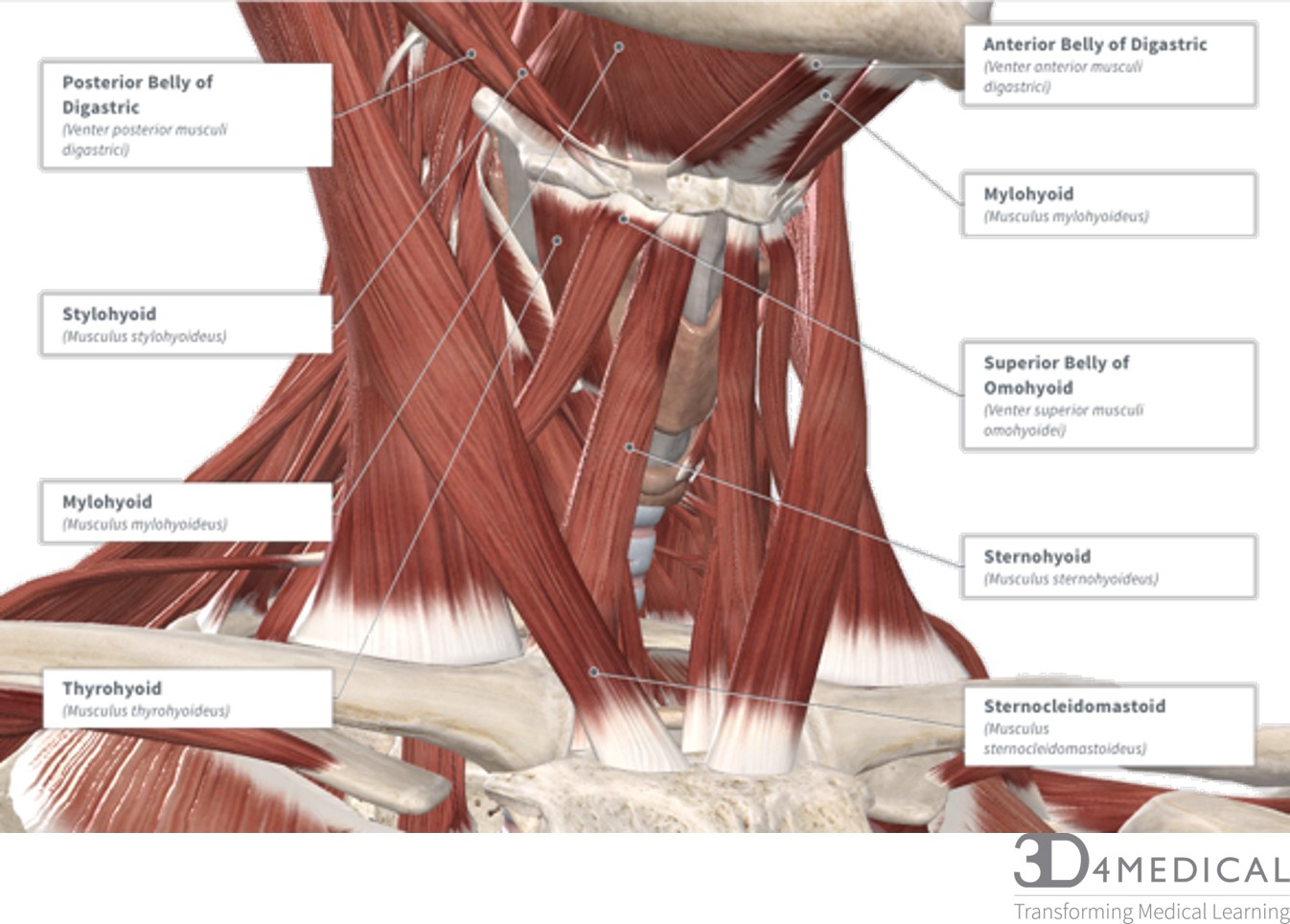
| Infrahyoid Muscles | |||||
| Muscle | Origin | Insertion | Action | Innervation | Blood Supply |
| Sternothyroid | Posterior surface of the manubrium inferior to the origin of the sternohyoid | Oblique line of the thyroid cartilage | Depression and stabilization of the larynx for pronation and swallowing | Superior root of ansa cervicallis and inferior root of ansa cervicallis | Cricothyroid branch of the thyroid artery and the lingual artery |
| Superior belly of Omohyoid | Intermediate tendon | Lower border of the hyoid bone lateral to the sternohyoid muscle | Depression and stabilization of the hyoid and larynx for swallowing and phonation | Superior root of ansa cervicallis | superior thyroid artery and lingual artery |
| Inferior belly of Omohyoid | Superior border of the scapula medial to the scapular notch | Intermediate tendon | Depression and stabilization of the hyoid and larynx for swallowing and phonation | Inferior root of ansa cervicallis | Superior thyroid artery |
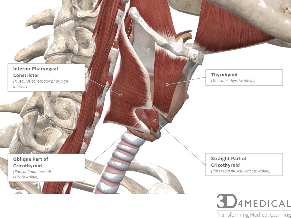
| Thyrohyoid | Oblique line of the thyroid cartilage | Lower border of the hyoid bone | Depression and stabilization of the hyoid and elevation of the larynx during swallowing | C1 spinal nerve | Superior thyroid artery and lingual artery |
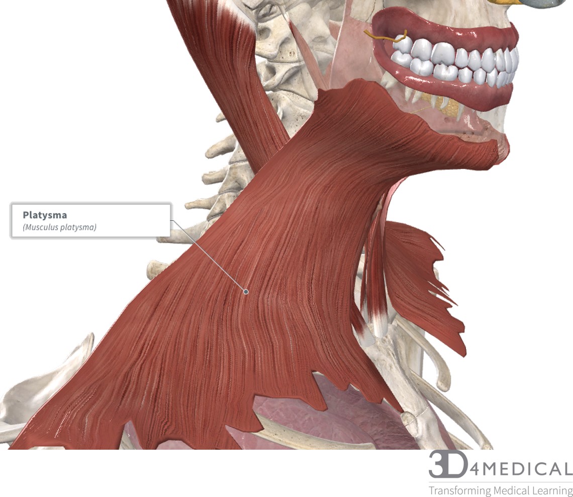
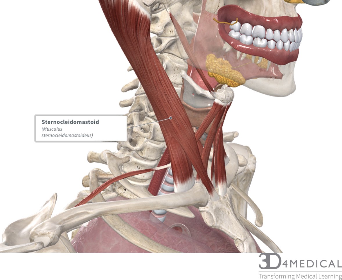
SUPERFICIAL ANTERIOR MUSCLES OF THE NECK
The two superficial muscles of the anterior neck, platysma, and sternocleidomastoid, contribute to 4 main actions: cervical spine flexion, lateral flexion of the cervical spine, cervical spine rotation, and depression/stabilization of the Mandible. The right and left sternocleidomastoid muscles can contract individually to rotate the cervical spine or flex laterally. When they contract together, neck flexion occurs.
| Muscle | Origin | Insertion | Action | Innervation | Blood Supply |
| Platysma | Pectoral fascia overlying the pectoralis major and deltoid muscles | Inferior border of the mandible and parotoid fascia | Depression of the mandible | Cervical branch of the facial nerve | Submental artery and suprascapular artery |
| Sternocledomastoid | Anterior surface of the manubrium, the medial third of the clavicle | Mastoid process and the lateral part of the nuchal lines | Cervical spine lateral flexion, cervical spine rotation and cervical spine flexion | Accessory nerve | Occipital artery, posterior auricular artery, superior thyroid artery, suprascapular artery |
MUSCLES SURROUND THE LARYNX
The muscles surrounding the larynx either insert or originate on the laryngeal skeleton or the hyoid bone. This means that these muscles aid in swallowing and positioning the vocal cords.
| Muscles surrounding the Larynx | |||||
| Muscle | Origin | Insertion | Action | Innervation | Blood Supply |
| Thyrohyoid | Oblique line of the thyroid cartilage | Lower border of the hyoid bone | Depression and stabilization of the hyoid and elevation of the larynx during swallowing | C1 spinal nerve | Superior thyroid artery and lingual artery |
| Inferior Pharyngeal Constrictor | Oblique line of the thyroid cartilage | Pharyngeal raphe | This muscle contracts to push the bolus of food downward into the esophagus | Pharyngeal branch of the vagus nerve, external branch of the superior laryngeal nerve | Ascending pharyngeal artery and the superior laryngeal artery |
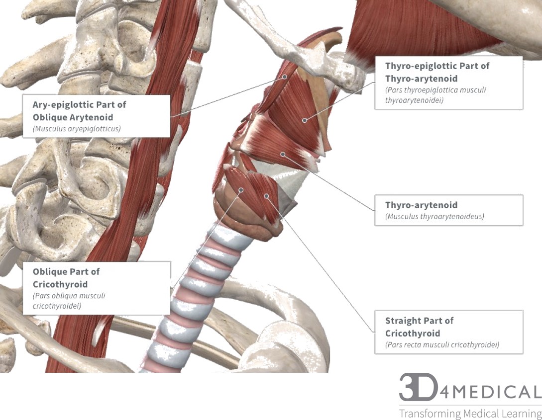
| Cricothyroid (Straight part) | Anterior surface of the cricoid cartilage, medial to oblique part | Inferior margin of the thyroid margin, inferior Cornu of the thyroid cartilage | Laterally rotates the thyroid cartilage and pulls it inferiorly to tighten the vocal cords | External branch of superior laryngeal nerve | External branch of superior laryngeal nerve |
| Cricothyroid (Oblique part) | Anterior surface of the cricoid cartilage, lateral to straight part | Inferior margin of the thyroid margin, inferior Cornu of the thyroid cartilage | Laterally rotates the thyroid cartilage and pulls it inferiorly to tighten the vocal cords which increases pitch | External branch of superior laryngeal nerve | Cricothyroid branch of superior thyroid artery |
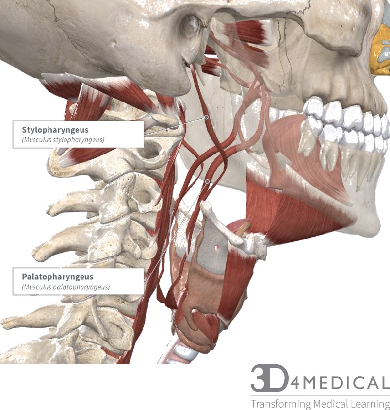
| Palato-pharyngeus | Hard palate, palatine aponeurosis | Posterior border of the thyroid cartilage, lateral wall of pharynx | Tenses the soft pallet and pulls the walls of the pharynx anteriorly, superiorly and medially | Pharyngeal branch of the vagus nerve, Pharyngeal plexus of the vagus nerve | Ascending palatine artery, greater palatine artery, ascending pharyngeal artery |
| Stylo-pharengeus | Anterior surface of the styloid process | Posterior border of the thyroid cartilage, muscular fibers of the superior and middle | Elevation of the pharynx and larynx | Glosso-pharyngeal nerve | Ascending palatine artery, tonsillar branch of facial artery, lingual artery, ascending pharyngeal artery |
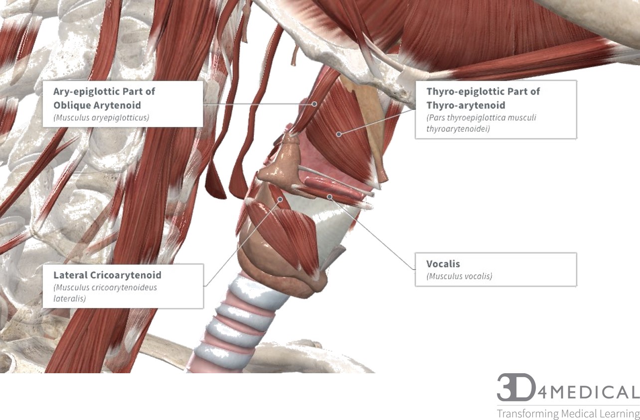
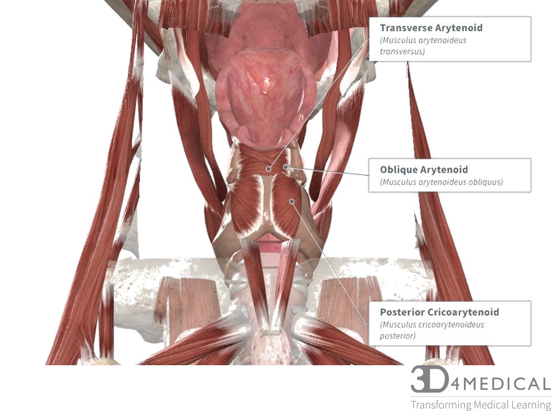
INTRINSIC MUSCLES OF THE THYROID CARTILAGE
The intrinsic muscles of the larynx are deep to the thyroid cartilage and the median thyrohyoid ligament. The actions of these muscles dictate the voice.
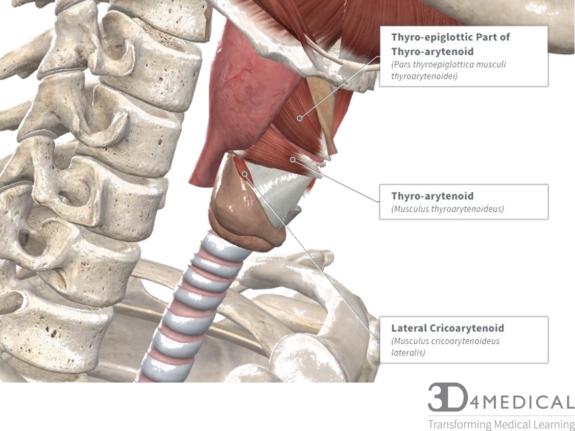
The internal and external thyro-artyenoid muscles change the vocal pitch by either contracting or relaxing. In isolation, the contraction of these muscles lowers the resonant frequency of the vocal folds.
| Intrinsic muscles of the Thyroid Cartilage | |||||
| Muscle | Origin | Insertion | Action | Innervation | Blood Supply |
| Lateral Cricioartyenoid | Lateral Part of the cricoid cartilage arch | Lateral Part of the cricoid cartilage arch | Opposes the action of the posterior cricioartyenoid by medially rotating and adducting the vocal cords | Inferior laryngeal nerve | Inferior laryngeal nerve |
| Posterior Cricioartyenoid | Posterior surface of the cricoid cartilage | Posterior muscular process of arytenoid cartilage | Opens the rima glottidis by pulling the vocal ligaments laterally and anteriorly; laterally rotates the arytenoid cartilage | Inferior laryngeal nerve | Superior laryngeal artery and inferior laryngeal artery |
| Transverse Arytenoid | Lateral border of the right and left posterior muscular processes of the arytenoid cartilage | Lateral border of the right and left posterior muscular processes of the arytenoid cartilage | Adducts the anterior portion of the arytenoid cartilage by pulling the posterior portion together | Inferior laryngeal nerve | Superior laryngeal artery and inferior laryngeal artery |
| Oblique Arytenoid | Posterior surface of the muscular process of arytenoid cartilage | Apex of the contralateral arytenoid cartilage and aryepiglottic fold | Closes the laryngeal inlet when swallowing or coughing | Inferior laryngeal nerve | Superior laryngeal artery and inferior laryngeal artery |
| Ary-epiglottic part of Oblique Arytenoid | Apex of the arytenoid cartilages on its respective side | Lateral margin of the epiglottis | Depresses the base of the epiglottis and closes the laryngeal inlet | Inferior laryngeal nerve | Superior laryngeal artery and inferior laryngeal artery |
| External Thyro-arytenoid | Thyroid angle of the posterior thyroid cartilage and the cricothyroid cartilage between the ary-epiglottic part of oblique arytenoid and the lateral cricioartyenoid muscles | Base and anterior surface of the arytenoid cartilage | Modulates vocal pitch | Inferior laryngeal nerve | Superior laryngeal artery and inferior laryngeal artery |
| Thyro-epiglottic part of Thyro-arytenoid | Internal surface of the thyroid cartilage | Margin of the epiglottis | Depresses the base of the epiglottis which closes the laryngeal inlet | Inferior laryngeal nerve | Superior laryngeal artery and inferior laryngeal artery |
| Internal Thyro-arytenoid (aka Vocalis) | Vocal process of arytenoid cartilage | Vocal ligament and thyroid cartilage | Modulates vocal pitch by Contracting or relaxing the vocal cords | Inferior laryngeal nerve | Superior laryngeal artery and inferior laryngeal artery |
POSTERIOR CERVICAL MUSCLES
Posterior muscles of the cervical spine primarily cause neck extension and assist in holding the head in an upright position and are often exercised in unison. All muscles have mirror images on the left and right side of the neck and when both are activated at the same time, the head and neck will move as indicated in the table below. However, when used separately, each muscle can move the head in a lateral motion (ipsilateral, same side of the body) with the angle depending on points of origin and insertion.
In the table below muscles are grouped by their location (superficial to deep, medial to lateral and superior to inferior) and function. The Trapezius (truh-pee-zee-uh-s) and Levator Scapula (see Figure below) are relatively superficial and both cause neck movement as well as the elevation of the scapula.
The trapezius is a vast muscle and extends from the skull to lower thoracic spine. Functionally the trapezius can be divided into Descending Portion (upper), Transverse Portion (central, laterally stretching) and Ascending Portion (lower). In the table below, we are looking at the Descending Portion of the Trapezius (pars descendens musculi trapezii), please click here for more on the central and lower portions.
When activated, the descending trapezius causes neck extension and hyper-extension, and with activation on one side, the descending trapezius can cause lateral neck flexion as well as shoulder elevation.
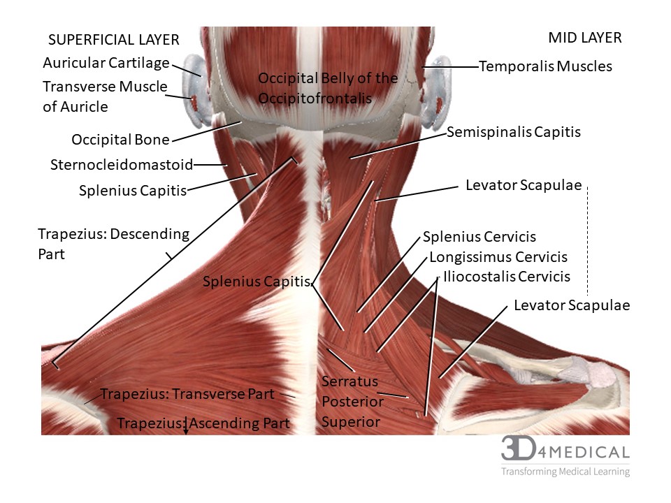 Figure. Posterior Cervical Muscles Superficial and Mid-Layers
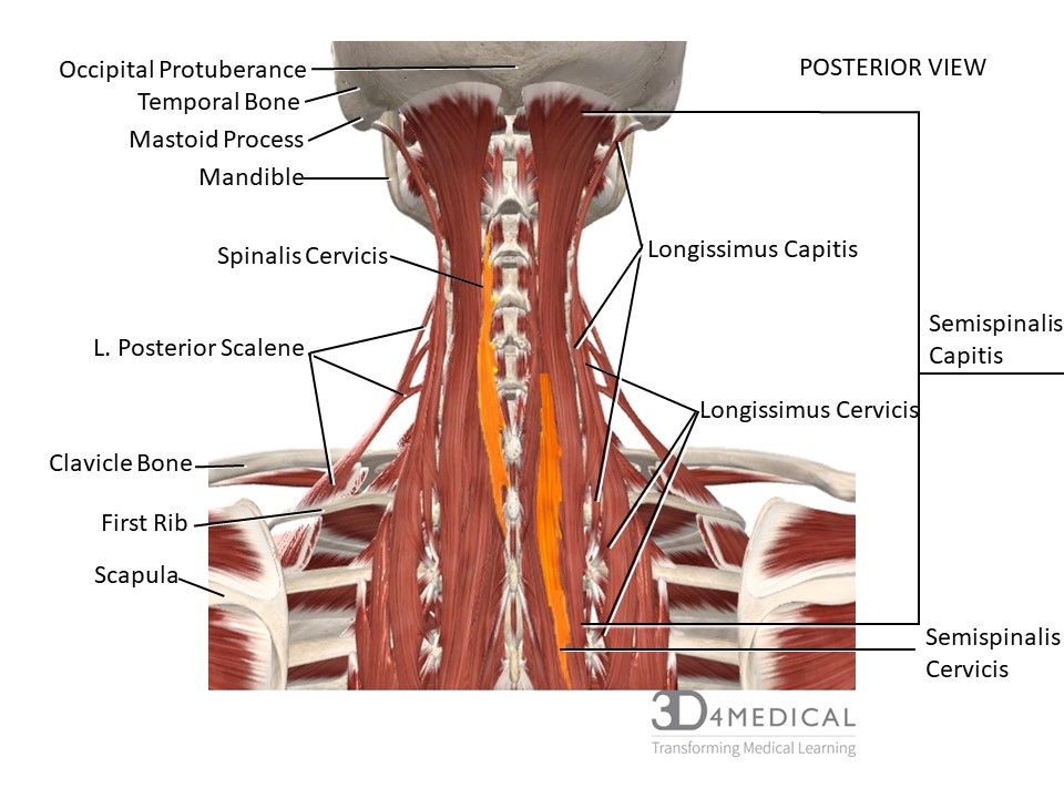
Figure. Posterior Cervical Muscles Deep Layers. (See Below for Spinal Muslces)
Table 1: Overview of Cervical Muscles from Superficial to deep, medial to lateral and superior to inferior. (For deep cervical spine muscles, click here); PDF File of Table

Below is an image giving perspective on how the many muscles of the head attach to the skull. Some of these groupings include muscles for movement of the head and neck, for mastication (chewing), and for some facial movements.
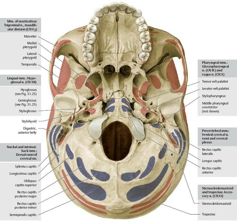
Figure X. Muscle attachment points on the inferior surface of skull, mandible removed (Norma basalis). Groupings of muscles with nerves noted. n.= nerve, m.= muscle (singular), mm.= muscles (plural)
Add to your learning of the muscles that move the head and neck: https://www.getbodysmart.com/neck-muscles
Deep muscles associated with the cervical region
Intervertebral muscles: The intervertebral muscles main function is to provide motion to the spine. Please refer to Table 1 for specific Origins, Insertions, Actions, and Innervations, of the intervertebral muscles.
Cervical interspinales: In the cervical region the interspinales muscles are the most distinct which consist of 6 pairs. They are found between the spinous processes of the vertebrae. The shape is also small narrow bundles. In the cervical region, they range from the inferior part of the axis’s spinous process down to the interspinales muscles between C7 and T1. In the thoracic region, they are found between T1-T2, T11-T12 and sometimes T2-T3. In the lumbar region, interspinales muscles are found between all the lumbar vertebrae, and sometimes between T-12-L1 and L5-S1.
Figure 7: Cervical Intervertebral muscles
Cervical intertransversarii: In the cervical region the intertransversarii muscles are small muscles placed between the transverse processes of vertebrae. These muscles also span most of the spinal column but do not appear between thoracic vertebrae T1-T9. These muscles have seven pairs. There is an anterior and posterior intertranserverarii between each transverse process and are separated by a cervical nerve that runs between each intertransversarii.
Mutifidus muscles: In the cervical region the multifidus muscles are long and thin muscles that are found on the articular processes of the bottom 4 vertebrae. The multifidus muscles span nearly the entire length of the vertebral column from the sacrum to the axis. They are very thin muscles that fill up the groove between the spinous processes of vertebrae. They play an important role in stabilizing joints within the spine. The multifidus muscles are bilateral in nature and span three joint segments.
Rotatores group: (rotatores brevis, longus, capitis, thoracis, and lumborum): In the cervical region that rotatores muscles run up and down the spine and lie beneath the multifidus muscles. The rotatores muscles include the rotatores capitis, rotatores cervicis (brevis and longus), and rotatores thoracis. At the cervical level and lumbar level, the rotatores may not be as obviously present and may be represented by a deeper bundle of tissues like the multifidus muscles. The Rotatores muscles are small and quadrilateral in shape and are also are involved in proprioception and postural control.
Deep suboccipital and anterior cervical muscles
Suboccipital muscles: The suboccipital muscles in the cervical region are named according to their position below the occipital bone. Their primary function is to stabilize the head and neck. Please refer to Table 2 below for specific Origins, Insertions, Actions, and Innervations of the suboccipital muscles and the deep anterior cervical muscles Longus Colli and Longus Capitis.
Rectus capitis posterior minor: The rectus capitis posterior minor is a postural muscle that acts at the atlanto-occipital joint and that monitors that position of the head. It is considered to be a sensory organ as well as a muscle that extends the head and neck.
Figure 8: Suboccipital cervical muscles
Rectus capitis posterior major: The rectus capitis posterior major muscle acts at the atlanto-occipital joint allowing for extension and rotation of the head. This muscle forms the superior part of the suboccipital triangle.
Obliquus capitis superior: The obliquus capitis superior muscle is a small muscle that forms the lateral part of the suboccipital triangle. It is deep to the trapezius
Obliquus capitis inferior: The obliquus capitis inferior muscles are the larger muscle in this group of two muscles. This muscle forms the inferior part of the suboccipital triangle. It is also deep to the trapezius and semispinalis capitis. Although the muscles named “capitis” it doesn’t actually connect to the cranium.
Deep anterior cervical muscles
Longus Colli: The longi colli muscles are made up of three parts (the superior oblique portion, the medial vertical portion, and the inferior oblique portion). It is found on the anterior part of each vertebra between the atlas and third thoracic vertebrae. This muscle is broad in the middle and narrow at either end. These muscles are often injured in rear whiplash accidents (This will be covered further in the common injury section).
Figure 9: Deep anterior cervical muscles
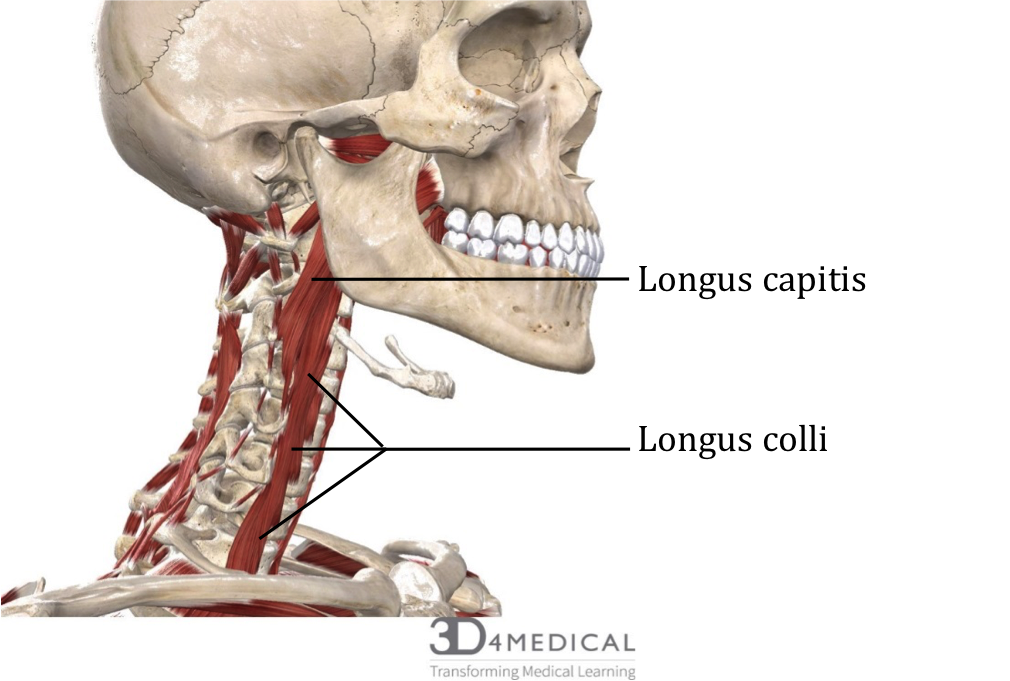
Longus capitis: The longus capitis is broad at the superior end and narrow below where it attaches to the tubercles of the anterior transverse processes.
Table 1: Cervical Spine Intervertebral Muscles
| Muscles | Origin | Insertion
|
Action | Innervation |
| Interspinales | Spinous processes of all vertebrae (Axis-T1) | Spinous process of next superior vertebrae | Extends back and neck | Dorsal Rami of (cervical) spinal nerves |
| Intertranversarii | Transverse process of each vertebrae | Transverse process of more superior vertebrae | Lateral flexion of vertebral column | Dorsal Rami of (cervical) spinal nerves |
| Multifidus | Articular processes of (C4-C7) | Spinous processes of superior vertebrae | Extends and rotates vertebral column | Dorsal Rami of (cervical) spinal nerves |
| Rotatores Cervicis
(Brevis) |
Posterior part of the transverse process for cervical | Base of the spinous process of above vertebra | Extends, flexes and rotates vertebral column and proprioception | Dorsal Rami of (cervical) spinal nerves |
| Rotatores Cervicis (Longus) | Superior part of transverse process | Lower border and lateral surface of the lamina of 2nd above vertebra | Extends, flexes and rotates vertebral column and proprioception | Dorsal Rami of (cervical) spinal nerves |
Table 2: Deep suboccipitals and deep anterior cervical muscles
| Muscles | Origin | Insertion | Action | Innervation |
| Rectus capitis posterior major | Spinous process of the axis (C2) | Inferior nuchal line of occipital bone | Ipsilateral rotation of head and extension | Dorsal ramus of the atlas (suboccipital nerve) |
| Rectus capitis posterior minor | Tubercle on posterior arch of atlas | Medial part of the inferior nuchal line of occipital bone | Extends head at neck & sensory organ | Suboccipital nerve |
| Obliquus capitis superior | Lateral mass of atlas | Lateral half of the inferior nuchal line | Extends head and flexes heads to the ipsilateral side | Suboccipital nerve |
| Obliquus capitis inferior | Spinous process of axis | Lateral mass of atlas | Rotations of head and neck | Suboccipital nerve |
| Longus capitis | Anterior tubercles of transverse processes of 3- 6 cervical vertebrae | Basilar part of occipital bone | Flex the head a neck laterally and bilaterally and rotates head ipsilaterally | C1-C4 |
| Longus colli | Transverse processes of the C5-T3 | Anterior arch of atlas | Flexes the head and neck | C2-C6 |
