Lower Limb
Muscles
Muscles of the Thigh
The thigh can be organized into five groups by the actions/location: the adductors, the lateral rotators, the gluteal group, the quadriceps, the hamstrings, and the iliopsoas.
Adductors
Pectineus originates at the pectineal line along superior ramus of pubis, inserting on the posterior surface of the femur and inferiorly on the lesser trochanter. Its actions cause flexion, medial rotation, and adduction at the hip. It is innervated by the femoral nerve (L2-L4).
Gracilis is the most superficial and medial of this muscle group. It crosses at both the hip and knee joints, making it a biarticulate muscle. It is sometimes surgically grafted into the hand or forearm to replace a damaged muscle. Gracilis originates from the inferior rami of the pubis and the body of the pubis. It inserts to the medial surface of the tibia, between the tendons of the sartorius (anteriorly) and the semitendinosus (posteriorly). Since it crosses the joint, it acts on to different parts of the leg; it causes adduction at the thigh and flexion at the knee. It is innervated by the obturator nerve (L2-L4).
Adductor Magnus is the largest muscle of the medial compartment, lying posteriorly of the group. It is commonly referred of having two parts; the adductor portion and the hamstring portion. The adductor portion originates from the inferior pubis ramus inserting onto the linea aspera of the femur. The hamstring portion originates from the ischial tuberosity and inserts onto the adductor tubercle and medial supracondylar line of the femur.
Actions of both portions include hip adduction, flexion, and extension. The adductor portion is innervated by the obturator nerve (L2-L4) while the hamstring portion is innervated by the tibial nerve (L4-S3).
Adductor Longus is a large, flat muscle, that partially covers adductor brevis and magnus and forms the medial border of the femoral triangle. Adductor longus originates from the pubis and expands into a fan shape, inserting onto the linea aspera of the femur. Actions include hip adduction and medial rotation of the thigh. Nerve innervations are by the obturator nerve (L2-L4).
Adductor Brevis is a short muscle, lying inferior to adductor longus. It originates from the inferior pubic rami and inserts on the posterior surface of the linea aspera. The action is adduction of the thigh. Nerve innervations are done by the obturator nerve (L2-L4).
Adductor Brevis originates on the inferior pubic ramus and inserts onto the linea aspera. Its action is to externally rotate the hip and plays a role in hip adduction as well. It’s innervated by the obturator nerve (L2-L4).
Gluteal Group
Gluteus Maximus is the largest muscle of the gluteal group. It originates on the iliac crest, the lateral surface of the ilium, sacrum, and coccyx. The insertion point is along the iliotibial tract and the gluteal tuberosity. On its own, the gluteus maximus has actions at the hip producing extension and lateral rotation. Its innervated by the inferior gluteal nerve (L5-S2).
Both gluteus medius and gluteus minimus, deep to gluteus maximus, share the same origin, insertion, action and nerve innervations. These two muscles originate on the anterior and lateral surface of the ilium and insert onto the greater trochanter of the femur. The actions produced are abduction and medial rotation at the hip. The nerve innervation is caused by the superior gluteal nerve (L4-S1).
Tensor fasciae latae (TFL) is a muscle that runs along continuous with the iliotibial tract which is the insertion point while it originates along the iliac crest of the anterior superior iliac spine. The TFL assists in keeping the balance of the pelvis while standing, walking or running and causes extension of the knee and lateral rotation of the leg. The nerve innervation is the superior gluteal nerve (L4-S1).
Lateral Rotators
Obturators externus is a muscle that originates along the lateral and medial margins of the obturator foramen and inserts onto the medial surface of the greater trochanter of the femur. Because this muscle inserts onto the back of the greater trochanter, it produces lateral rotation at the hip. The nerve innervation is by the obturator nerve (L3-L4).
Obtruator internus is the partner to the obturator externus that originates similarly along the lateral and medial margins of the obturator foramen, however, the insertion differs with the insertion. Obturator internus inserts onto the inner surface of the greater trochanter, producing the action of lateral rotation at the hip. The nerve innervation is by the obturator nerve (L3-L4).
Piriformis is the other dominant muscle of the lateral rotator groups alongside the obturators. It originates on the anterolateral surface of the sacrum and inserts onto the greater trochanter of the femur. Likewise, to the name it produces lateral rotation at the hip and is innervated by the branches of the sacral nerves (S1-S2).
Gemellus superior and inferior is a pair of lateral rotators that both originate along the ischial spine and insert onto the medial surface of the greater trochanter of the femur. These two muscles produce lateral rotation at the hip and are innervated by the obturator internus and quadratus femoris nerves.
Quadratus femoris, is a flat quadrilateral muscle that originates along the lateral border of the ischial tuberosity. The insertion point is at the intertrochanteric crest of the femur and produces lateral rotation at the hip. The nerve innervations are by the sacral plexus (L4-S1).
Iliopsoas
Psoas major is a large muscle of the pair and originates on the anterior surfaces and transverse processes of the vertebrae. The insertion point of this muscle is at the lesser trochanter and produces flexion at the hip. The nerve innervation is caused by the branches of the lumbar plexus (L2-L3).
Illiacus, the partner to psoas major, originates along the iliac fossa of the ilium and the insertion point is the merging of the tendon with psoas major. The action produced is flexion at the hip and innervated by the femoral nerve (L2-L3).
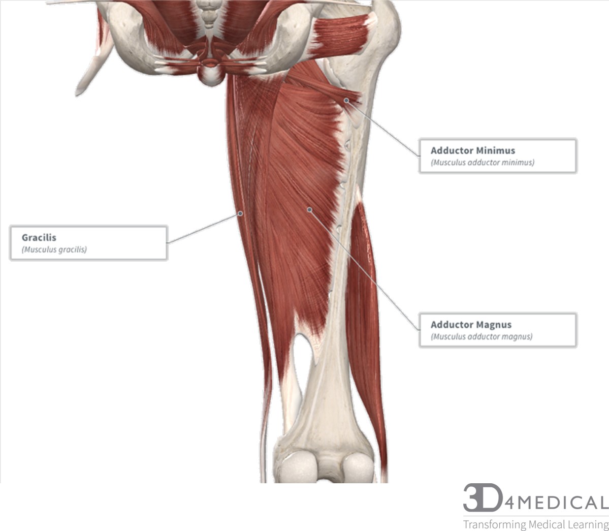
Figure 1. Diagram representing the posterior view of the thigh showing the adductor magnus, gracilis and adductor minimus muscles.
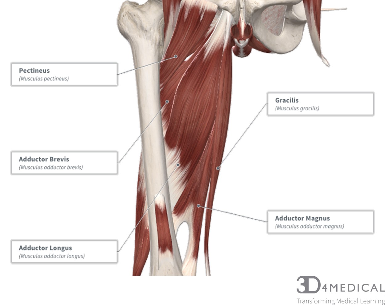
Figure 2. Diagram representing the anterior view of the muscle groups adductor brevis, adductor longus and adductor magnus.
Hamstrings
Biceps femoris muscle has two (Bi-) heads, the long head, and the short head. It is the most lateral of the muscles of the group; the common tendon of the two heads can be palpated laterally at the posterior knee. The long head originates from the ischial tuberosity of the pelvis. The short head originates from the linea aspera on the posterior surface of the femur. Together, the heads form a tendon, which inserts into the head of the fibula. The main action is flexion at the knee. It also causes extension and lateral rotation of the hip. The long head is innervated by the tibial part of the sciatic nerve, whereas the short head is innervated by the common fibular portion of the sciatic nerve (L5-S2).
Semitendinosus is a largely tendinous muscle. It lies medially to the biceps femoris and is superficial to semimembranosus. It originates from the ischial tuberosity of the pelvis and inserts to the medial surface of the tibia. Its actions are flexion at the knee, extension and medial rotation of the hip. It’s innervated by the tibial portion of the sciatic nerve (L5-S2).
Semimembranosus is a flatter, broad shaped muscle. It is located inferiorly to the semitendinosus muscle. It originates from the ischial tuberosity and inserts on the medial tibial condyle. Its actions are knee flexion with extension and medial rotation at the hip. It’s innervated by the tibial portion of the sciatic nerve (L5-S2).
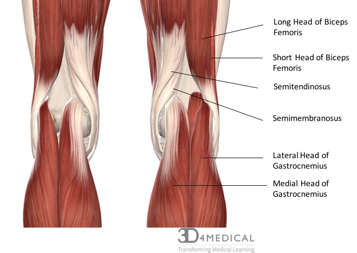
Figure 3. Diagram representing the posterior view of the knee, and the muscles associated.
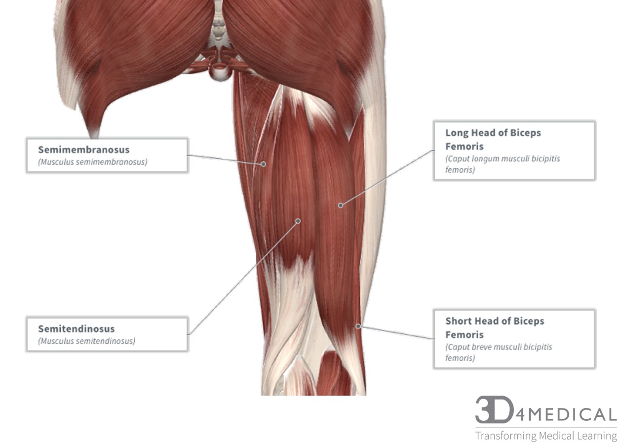
Figure 4. Diagram represnting the posterier view of the hamstring muscle group.
Quadriceps
Rectus Femoris, named for its muscle fascicle orientation (rectus = straight) is the most superficial muscle on the anterior compartment. This muscle is primary mover in knee extension and assists with hip flexion. This muscle originates on the anterior inferior iliac spine and inserts onto the tibial tuberosity via the patellar ligament/quadriceps tendon. Nerve innervation via the femoral nerve (L2-L4).
Vastus Lateralis is a large (vast = large) lateral muscle. The origin is on the anterior and inferior surface of the greater trochanter and proximal linea aspera which inserts onto the tibial tuberosity via the patellar ligament/ quadriceps tendon. Its main action causes knee extension. Nerve innervation via the femoral nerve (L2-L4).
Vastus Intermedius is deep to rectus femoris. This muscle originates on the anterior lateral shaft of the femur and distal lines aspera and inserts onto the tibial tuberosity via the patellar ligament/quadriceps tendon. Its main action causes knee extension. Nerve innervation via the femoral nerve (L2-L4).
Vastus Medialis is located along the medial portion of the anterior compartment. It originates along the entire length of the lines aspera and inserts onto the tibial tuberosity via the patellar ligament/quadriceps tendon. The main action causes knee extension. Nerve innervation via the femoral nerve (L2-L4).
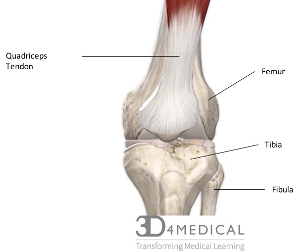 Figure 5. Diagram representing the anterior view of the quadriceps tendon inserting over the patella.
Sartorius is a unique muscle because it is the only knee flexor that originates anteriorly. Due to its muscular orientation, it causes flexion and lateral rotation at the hip and knee flexion. It originates is on the anterior superior iliac spine on the lateral portion of the hip and crosses to insert medially on the tibia. Since this muscle crosses both the hip joint and the knee joint it is considered a biarticulate muscle, strictly meaning Bi (two) articulate (joints).
Articularis Genus is superior to the patella and superficial to the quadriceps tendon. Articularis Genus pulls the suprapatellar bursa superiorly during extension of the knee and prevents impingement of the synovial membrane between the patella and the femur.

Figure 6. Diagram representing the anterior view of the quadirceps mucle group.
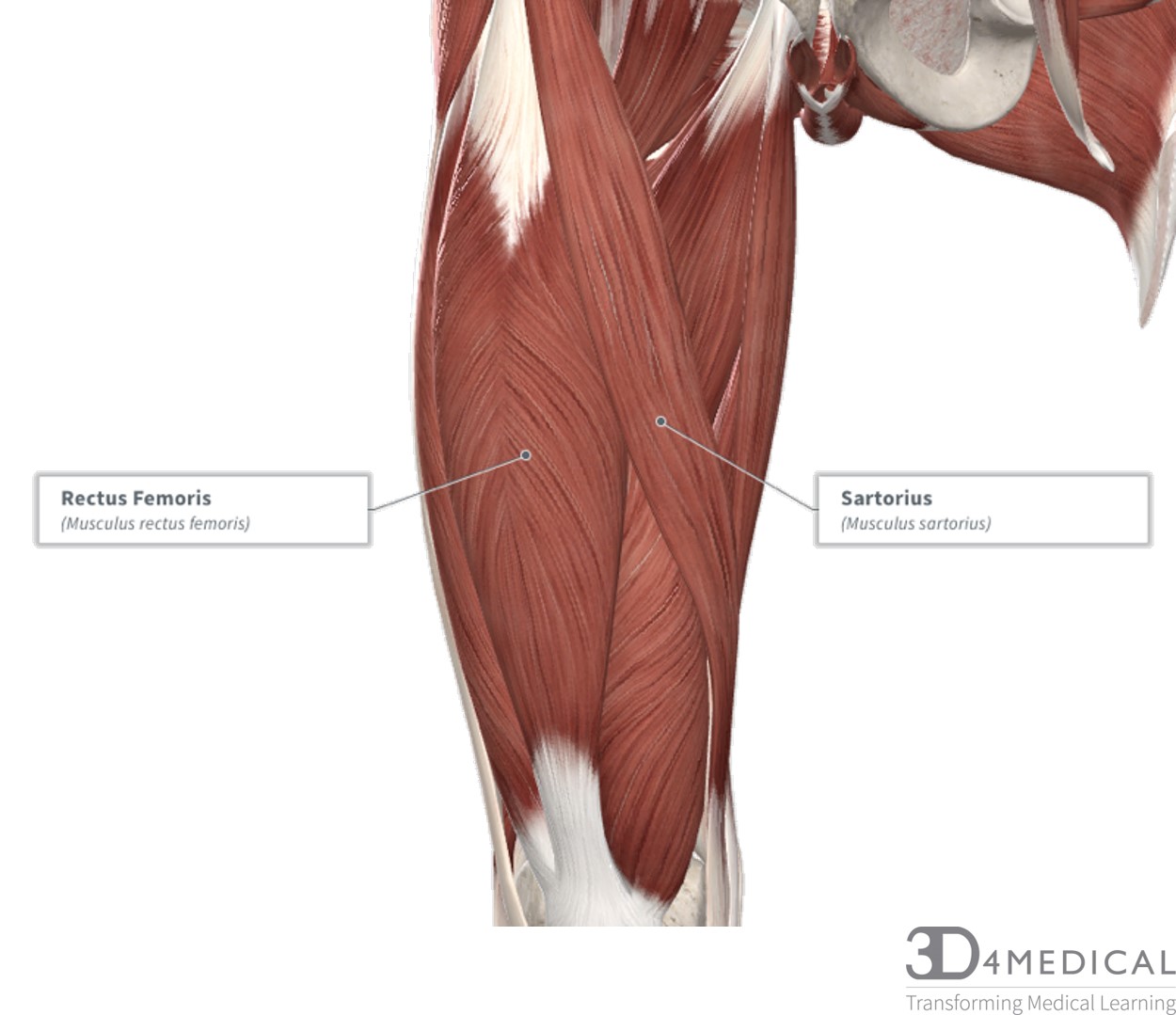
Figure 7. Diagram demonstrating a close-up, anterior view of the deep quadriceps muscles sartorius and rectus femoris
To challenge yourself or learn with some quizzes on the muscles visit: https://www.getbodysmart.com/anterior-thigh-muscles
Muscles in the Knee
Popliteus is a small muscle which is located on the posterior side of the knee. It originates on the lateral condyle of the femur and inserts on the posterior surface of the tibia above the soleus. Popliteus plays a role in both flexion and internal rotation of the knee. The arcuate popliteal ligament, which extends from the fibular head to the lateral condyle of the femur helps to secure popliteus in its place.
Plantaris is a thin rope-like muscle, with its tendon running down the posterior portion of the lower leg parallel to the much larger calcaneal or Achilles Tendon. Plantaris originates on the lateral condyle of the femur just superior and medial to the lateral head of gastrocnemius. Its insertion point is on the posterior side of the Calcaneus via the calcaneal tendon. The action of plantaris is to work with the calcaneal tendon to plantarflex the foot as well as aid in flexion of the knee.
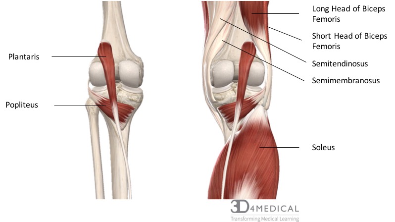
Figure 8. Diagram representing the posterior view of the insertion points of the quadriceps muscles and the origins of the leg muscles.
Muscles of the Leg
Anterior Compartment
Tibialis Anterior originates along the lateral shaft of the tibia and inserts onto the base of the first metatarsal bone and medial cuneiform. The action produced causes dorsiflexion of the ankle and inversion of the foot. The nerve innervation is the deep fibular nerve (L4-S1).
Extensor Digitorum Longus originates on the head of the fibular and inserts on the base of proximal phalanges 2-4. It causes extension of the 2nd, 3rd, and 4th digits and is innervated by the fibular nerve (L4-S1).
Extensor Hallicus Longus originates on the anterior surface of the tibia and inserts into the distal phalanges 2-5. The action produced is the extension of the toes 2-5 and is innervated by the deep fibular nerve (L4-S1).
Fibularis Tertius originates along the distal anterior surface of the fibula and the interosseous membrane. The insertion point is along the dorsal surface of the 5th metatarsal and is innervated by the deep fibular nerve (L5-S1).
Posterior Compartment
Superficial
Gastrocnemius originates as two split heads on the femoral condyles and merges to insert into the calcaneal tendon. The action produced is plantar flexion of the ankle, inversion of the foot and flexion of the knee. The nerve innervation is by the tibial nerve (S1-S2).
Soleus is flat muscle, deep to the gastrocnemius and originates at the head of the head of the fibula. It as well inserts into the calcaneal tendon and is innervated by the sciatic nerve (S1-S2).
Deep
Tibialis Posterior originates on the proximal 2/3 of tibia and fibula and inserts onto the medial cuneiform and navicular. Its action causes plantar flexion and inversion of the foot and is innervated by the tibial nerve (S1-S2).
Flexor Digitorium Longus originates on the posterior surface of the tibia and inserts on the base of the distal phalanges 2-5. It causes flexion of phalanges 2-5 and is innervated by the tibial nerve (L5-S1).
Flexor Hallucis Longus originates on the inferior 2/3 of the posterior fibula and inserts on the base of the distal phalanx of the hallux. It causes flexion of the hallux and is innervated by the tibial nerve (L5-S2).
Lateral Compartment
Peroneal Longus and Brevis
Peroneal longus originates at the head of the fibula and crosses inferiorly to insert laterally of the 1st metatarsal. While peroneal brevis originates inferior 2/3 of the lateral fibula and inserts on the dorsal surface of the 5th metatarsal. Innervated by the superficial peroneal nerve (L5, S1, S2).
