The Head and Neck
Bones
The Skull
Skull, head, cranial vault, dome, noodle, and thinker are all words that are used to describe the same thing: the cranium. The skull is part of the axial skeleton and is divided into the two sections: the facial bones and the cranium. It is comprised of the cranium, which is round and houses the brain, and the facial bones which form the upper and lower jaws, the nose, orbits, and other facial structures. The adult skull is made up of 22 bones; 21 of them are immovable and the 22nd one is the mandible, the lower jaw, and is the only moveable bone of the skull. The cranial bones form the cranial cavity which holds and protects the brain.
The cranium is composed of eight bones: the frontal bone, two parietal bones, two temporal bones, the occipital bone, the sphenoid bone, and the ethmoid bone. A suture is an synarthrosis joint, an immoveable joint, between the bones and is how the bones of the cranium are connected. They are fibrous joints. There are four major sutures that connect the bones of the cranium together: the frontal or coronal, the sagittal, the lambdoid, and the squamous. The frontal suture connects the frontal bone to the two parietal bones. The sagittal suture connects the two parietal bones. The lambdoid connects the two parietal bones to the occipital bone. The squamous sutures connect the parietal bones to the temporal bones.
The Frontal Bone
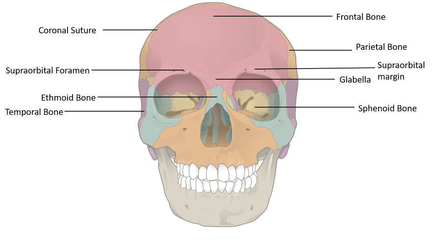
The frontal bone, as seen below in pink, is the anterior roof of the skull, the bone of the forehead, and extends down to be the superior portion of the orbits, the top of the eye sockets. It articulates (is connected to) with the parietal, sphenoid, ethmoid, nasal, lacrimal, maxillary, and zygomatic bones. The shape of the frontal bone plays a big part in the visual identity of a person. The supra-orbital margin is the superior, upper, rim of the eye socket. It has supra-orbital foramina, sometimes a notch, above each supra-orbital margin that allow blood vessels and nerves to reach the eyebrows, eyelids, and frontal sinuses. Supra meaning superior, orbital meaning eye socket, and foramen meaning hole; the name of the describes what it is and where it is. The smooth part above and between your eyebrows has a name and is called the glabella. The zygomatic process of the frontal bone connects with the frontal process of the zygomatic bone which help form the cheekbone.
The Parietal Bone
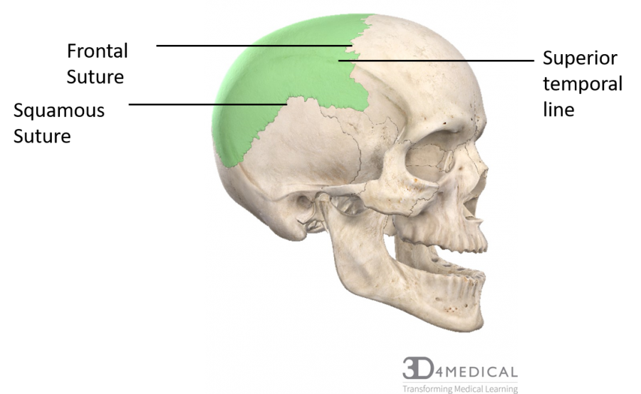
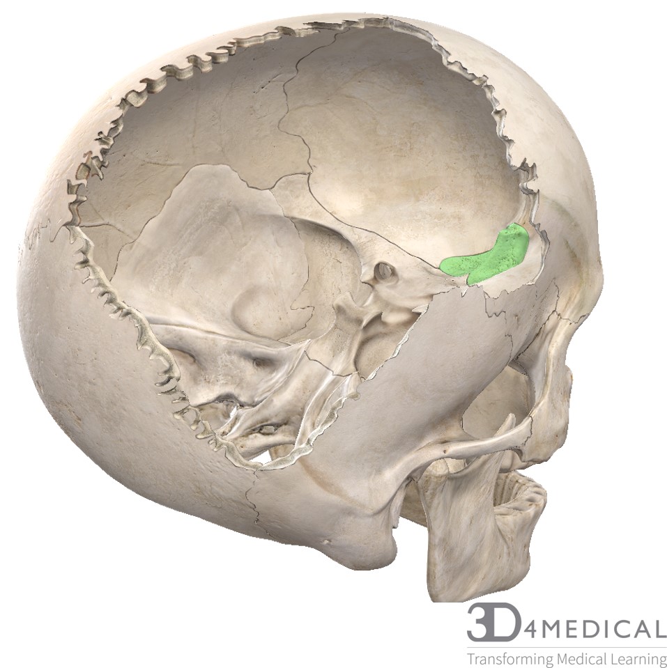
The parietal bones, the right parietal bone highlighted in green below, make the roof of the cranium. It forms the upper lateral side of the skull. The parietal bones articulate with one another, and with the frontal, sphenoid, temporal, and occipital. The superior and inferior temporal lines are low ridges that mark the attachment sites of the temporalis muscle.
The Temporal Bone
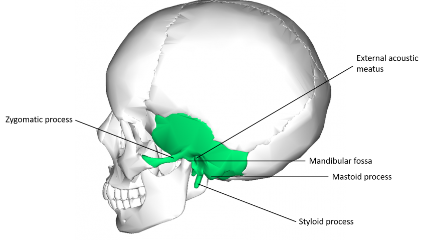
The temporal bones are the lower lateral sides of the skull as seen below in green. They are the bones one’s ears sit upon. There are many features of the temporal bones. First, there is the zygomatic process. It connects to the temporal process of the zygomatic bone. Together these form the zygomatic arch, the cheekbone. Next, there is the mastoid process. It is a pyramidal projection at the back of the temporal bone. It can easily be felt on the side of one’s head, behind the earlobe. It is a site of muscle attachment for muscles including sternocleidomastoid, splenius capitis, and longissimus capitis. The mandibular fossa of the temporal bone is a depression that articulates with the mandible; they form the temporomandibular joint. This allows for the opening and closing of the jaw. The temporal fossa is a depression in the temporal region. It is one of the largest landmarks of the skull. The frontal bone, sphenoid bone, parietal bone, and temporal bone all contribute to the concavity of the fossa. The temporal lines are the superior border and the zygomatic arch is the inferior border. It is a site of muscle attachment for temporalis, a mastication (chewing) muscle. Next, there is the styloid process. It is posterior to the mandibular fossa. It is a pointy, stylus like process that is just below the ear. It is an attachment point for muscles and tendons of the neck and tongue. The external auditory or acoustic, meatus or canal, is the canal the funnels sound into the eardrum. It is the large opening on the lateral side of the skull often associated with the ear. It is a tube running from outer ear to the middle ear. The internal auditor, or acoustic, meatus or canal is a bony canal within the petrosal portion of the temporal bone that allows nerves and vessels from within the posterior cranial fossa to the brain. It connects the middle and inner ear cavities to the temporal bone.
The Occipital Bone
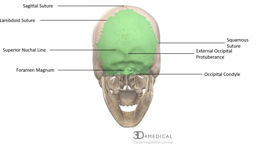
The occipital bone, highlighted in green below, is the bone the makes up the posterior and base portion of the cranium. The large oval opening in the base is the foramen magnum. It is where the spinal cord exits the skull. There are two oval shaped occipital condyles, one on each side of the foramen magnum. These are where the skull connects to the first cervical vertebrae of the vertebral column, atlas. At the midline of the bone, there is a protrusion called the external occipital protuberance, the tip of it is referred to as the inion. It is easy to find and palpate. This can vary in size in people. It is a site of attachment for a ligament of the posterior neck. Lateral to either side of the protrusion is the superior nuchal line. This is the most superior site for muscle attachment, with only the scalp covering the skull above the lines. Just inferior to the external occipital protuberance there is the inferior nuchal line, also a site of muscle attachment.
The Sphenoid Bone
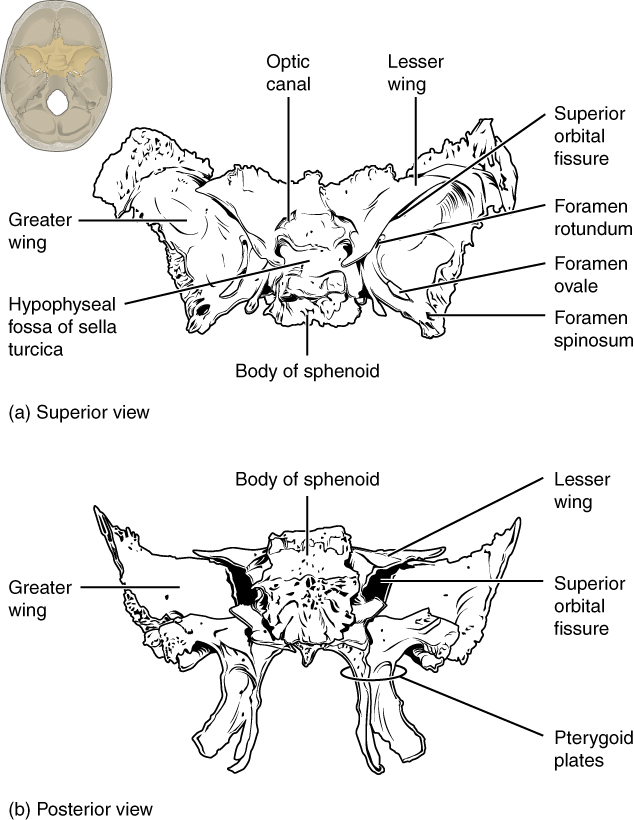
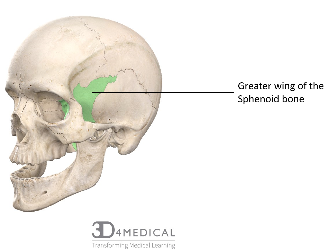
The Sphenoid bone, above, is a cranial bone shaped like a butterfly. It is located in the anterior portion of the cranium, as seen below in green. It forms the base of the anterior skull and spreads laterally enough to contribute to the sides of the skull. It consists of a central body that is cubical-shaped and two sets of wing-shaped extensions that project laterally: the greater wings and the lesser wings. The sella turcica (Turkish saddle) is located at the midline of the middle cranial fossa. It is named for the resemblance to a horse’s saddle. The hypophyseal (pituitary) fossa is the depression of the base of the sella turcica and houses the pituitary gland. The greater wings, also known as Ala major, extend out laterally from the sella turcica to form the anterior floor of the middle cranial fossa. They are a pair of larger winged structures of the bone, curved upwards, backward and on the lateral sides. The lesser wings, also known as Ala minor, are a pair of the smaller projections that are flat, triangular structures situated anterior to the greater wings. Projecting inferiorly on the posterior side are the media pterygoid plate and lateral pterygoid plate. The right and left medial pterygoid plates form the posterior, lateral walls of the nasal cavity. The larger lateral pterygoid plates serve as attachment sites for chewing muscles.
The Ethmoid Bone
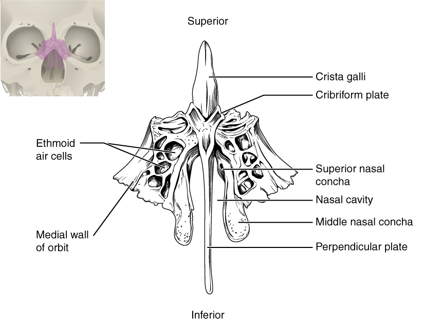
The ethmoid bone is located between the two orbits and at the roof of the nose. It forms the roof and lateral walls of the upper nasal cavity, the upper portion of the nasal septum, and contributes to the medial wall of the orbit. What makes it a cranial bone is that inferiorly it forms a portion of the anterior cranial floor. It separates the nasal cavity from the brain. It is made up of the perpendicular plate, the crista galli (rooster’s comb or crest), and the cribriform plate, as seen above. The perpendicular plate forms the superior portion of the nasal septum. The ethmoid bone also forms the lateral walls of the upper nasal cavity, extending from each lateral wall are the superior nasal concha and middle nasal concha, which are thin, curved projections that extend into the nasal cavity. The crista galli is a superior bony projection that serves as an attachment point for a covering layer of the brain. On either side of the crista galli is the cribriform plate. It forms the roof of the nasal cavity and contributes to the anterior cranial fossa. It is a small, flattened area with many small openings, olfactory foramina. Small nerve branches from the olfactory areas of the nasal cavity pass through these openings to enter the brain. You can see it highlighted in green below from a superior lateral view through the cranium.
Cranial Cavity
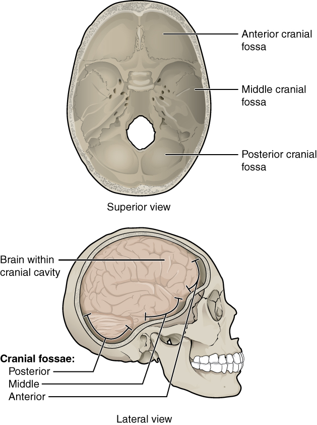
The inner surface of the cranial cavity is divided into three distinct depressions. It is comprised of the anterior, middle, and posterior cranial fossae. Each fossa accommodates a different part of the brain. The anterior cranial fossa is the most shallow and superior of the three fossae. The fossa is comprised of the frontal bone, the ethmoid bone, and the body and lesser wings of the sphenoid bone. It houses the anteroinferior portion of the frontal lobe. The middle fossa is made up of the greater wings of the sphenoid bone and the temporal bone. It can be said to be butterfly shaped. It houses the pituitary gland and the two lateral parts of the fossa contain the temporal lobes. The posterior cranial fossa is made up of mainly the occipital bone and the posterior section of the temporal bones. It houses the brainstem and cerebellum as well as associated arteries and nerves.
A mnemonic to remember the bones of the cranium is PEST OF.
| Mnemonic | Bones |
| P | Parietal |
| E | Ethmoid |
| S | Sphenoid |
| T | Temporal |
| O | Occipital |
| F | Frontal |
To see more about the bony landmarks of the skull which are used for radiological or anthropological skull measurements see: https://radiopaedia.org/articles/skull-landmarks
This video describes the bones of the cranium and face and shows them on a model. From 3:50 onward are the cranial bones. https://www.youtube.com/watch?v=0N20150qthA
Hyoid
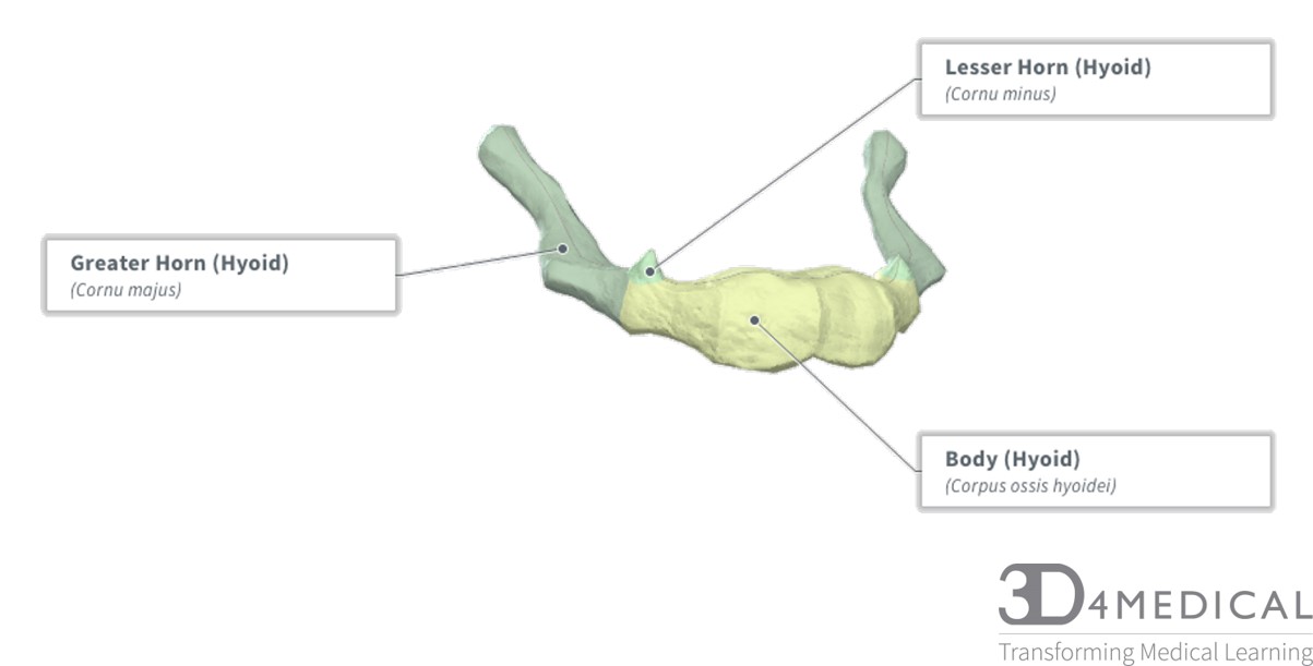
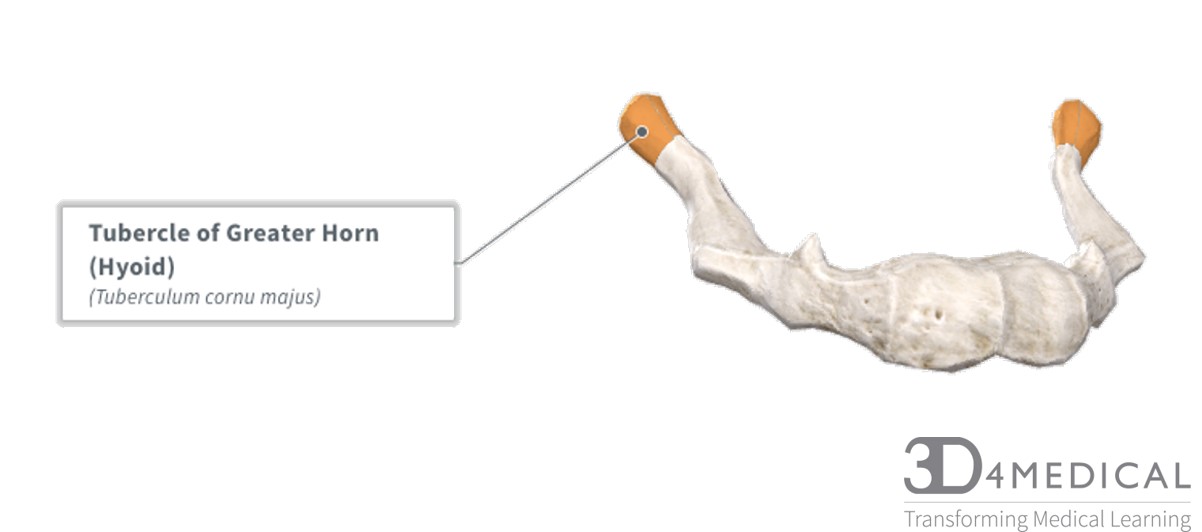
This horseshoe-shaped bone is used as a solid foundation for the tongue to move. This bone also has many areas for muscle attachment. The muscles above the hyoid are referred to as the suprahyoid muscles. The muscles below the hyoid are referred to as the infrahyoid muscles. For more information on these muscles, refer to the neck muscles section. There are 3 main parts of the hyoid: the body, the greater horn, and the lesser Horn. There is one major feature which is the tubercle of the greater horn.
The cervical spine region
Introduction:
The cervical spine is part of the axial skeleton and consists of the top seven vertebrae, which attaches the most superior vertebrae (C1 or atlas) to the base of the skull and connects the seventh cervical vertebra (C7) to the first thoracic vertebra (T1). The seven cervical vertebrae make up the neck of the body and hold up the head portion of the body. Along with holding the skull in place, these vertebrae play a role in allowing motion of the head and neck. Other functions of the cervical spine region also act as a site of attachment for many muscles and ligaments, as well as work as a protective structure for the spinal cord, spinal nerves, and cardiovascular system.
Additional cervical spine purposes act to house the superior portion of the spinal cord, which exits the head at the foramen magnum at the inferior part of the cranium through the occipital bone, which is a considered a cranial bone. The joint that holds the head to the cervical spine is called the atlanto-occipital joint, which allows some articulation at a synovial joint. The joint below the atlanto-occipital is named the atlanto-axial joint, which is a connection of vertebra C1 (Atlas) and C2 (Axis). This atlanto-axial joint is considered a complicated triple joint with both pivot and gliding joints. These joints will be discussed in further detail ahead.
Other cervical vertebrae features include intervertebral discs, which lie in between each vertebra in the vertebral column. Each intervertebral disc acts as a fibrocartilaginous joint and is made of the outer fibrous ring named the annulus fibrosus, and the inner material is named the nucleus polposus, which is made up of a gel-like center. These structures will also be discussed in greater detail as the sections ahead.
The cervical spine vertebrae
Figure 1: Lateral view of seven cervical vertebrae
The cervical vertebrae are specifically known for having small bodies; most have bifid spinous processes and all have transverse foramina in each transverse process through which vertebral arteries, veins, and sympathetic nerves travel through. The cervical spine also has “special” features for the two superior vertebrae C1 and C2 (Atlas and Axis) and C7 (the most inferior vertebra). One unique feature of the atlas and axis allows for specialized functions that allow the thicker superior section of the spinal cord to travel through each vertebra. Other special features of the atlas allow for connection to the base of the skull forming an ellipsoid synovial joint. The atlas and axis also have specialized structures, which allow the motion of “yes” from the atlas and occipital joint called the atlanto-occipital joint and “no” from the atlas and axis named the atlanto-axial joint. These unique vertebrae will also be covered in greater detail in their specific sections.
Anatomical boney features/landmarks
The cervical vertebrae are named C1-C7 from superior to inferior with the C1 being named the “Atlas” and C2 named the “Axis”. C1, C2, and C7 are considered “special” (irregular) cervical vertebrae and C3-C6 are often referred to as “regular” cervical vertebrae
Unique boney features of the cervical vertebrae:
- Body: absent in atlas (C1), C2 has the “dens” as a small body, small body in others compared thoracic and lumbar bodies
- Vertebral foramen: large vertebral foramen (largest part of the spinal cord). The shape is triangular in C2-C7
- Transverse process: have transverse foramina for vertebral arteries and veins
- Transverse foramina: house and protect the vertebral arteries and veins
- Spinous process: absent in C1, bifid in others, except C7, C7 also has a prominent spinous process compared to superior cervical vertebrae
- Articular facets: face superior and inferior
Primary functions of the cervical vertebral column
- Protection: encloses and protects the spinal cord within the spinal canal. Transverse foramina also house and protect vertebral arteries and veins
- Support: carries the weight of the head and superior vertebrae
- Axis: forms the central axis of the body and acts as a pivot point for the superior Atlas
- Movement: has roles in both posture
- and movement
- Vertebral body: Acts as a protective structure and transfers weight along the spinal column
- Vertebral foramen: houses and protects the spinal cord
- Transverse process: have transverse foramina that house and protect vertebral arteries, veins, and nerves
- Spinous process: acts as a landmark for surface anatomy and is an attachment site for muscles
- Articular facets: allows for articulation between 2 opposing vertebrae
Figure 2: Posterior view of Cervical Vertebrae 
“Irregular” first cervical vertebra (C1) is named the Atlas.
Figure 2: Superior view of Atlas (C1)

Unique boney features: The atlas has no body and no spinous process, the largest vertebral foramen, no transverse foramina, an anterior vertebral arch with pedicles, a posterior arch with laminae, transverse processes, a facet for the axis’s (C2) unique odontoid (also called the dens), the transverse ligament of atlas which holds the axis’s dens in place, inferior articular facets, and large superior articular facets.
Figure 3: Lateral view of Atlas (C1)

Functions: The atlas vertebra is a “special” cervical vertebra that attaches to both the skull to the spine making the atanto-occiptial joint and the second cervical vertebrae making the atlanto-axial joint. This joint allows for the motion of “yes.” The superior and inferior facets of vertebrae meet one another at smooth articular facets of each articular process. The atlas’s superior facets meet the occipital condyles for articulation with the occipital bone on the base of the skull. The vertebral foramen of inferior vertebrae combine to create the vertebral canal, which is where the spinal cord is housed. The atlas’s vertebral foramen has both the anterior and posterior arch, which protects the spinal cord. The atlas also is a site for many muscles attachments, which will be covered in the muscles section.
Figure 3: Posterolateral view of Atlas and Axis (Atlanto-axial joint)
“Irregular” second cervical vertebra (C2) is named the Axis.
Figure 4: Superior view of Axis (C2)
Unique boney features: The axis has an irregular small body, a bifid spinous process, vertebral foramen, pedicles, a posterior arch with laminae, transverse processes and foramina, inferior articular facets, and superior articular facets, a facet for the axis’s (C2) unique odontoid (also called the dens), the dens projects superiorly from anterior part of axis,
Figure 5: Lateral view of the axis

Functions: The Axis is a “special” cervical vertebra that attaches to the atlas creating the atlanto-axial joint as well as inferior vertebra C3. This joint is known for the motion “no.” The “dens” sits in the vertebral foramen of the atlas while the atlas rotates around it. The vertebral foramen of adjacent vertebrae combine to create the vertebral canal. The axis’s vertebral foramen has both the anterior vertebral body (the dens in this case) and the posterior arch which protects the spinal cord. The transverse foramina also act to house and protect the vertebral arteries, veins, and sympathetic nerves. The axis is also a site for many muscles attachments, which will be covered in the muscles section.
“Regular” cervical “vertebrae C3-C6
Unique boney features include: C3-C6 are “regular” vertebrae with a posterior arch, large vertebral foramen, small body, pedicles, laminae, bifid spinous processes, transverse processes and foramina, and superior and inferior articular facets.
Functions: C3-C6 are considered “regular” cervical vertebra that articulate with the vertebrae that are found superior and inferior to each specific vertebrae. The vertebral foramen of adjacent vertebrae combine to create the vertebral canal. Each vertebral foramen has both the anterior vertebral body and posterior arch which protects the spinal cord. The transverse foramina also act to house and protect the vertebral arteries and veins. C3 is also a site for many muscles attachments, which will be covered in the muscles section.
Cervical vertebrae C4 “Regular”
Figure 6: Superior view of C4 Unique boney features: C4 is a “regular” vertebra with a posterior arch, large vertebral foramen, small body, pedicles, laminae, bifid spinous processes, transverse processes and foramina, and superior and inferior facets.
Functions: C4 is a “regular” cervical vertebra that articulates with the superior C3 and inferior C5. C4’s functions are similar to C3-C7 with many muscles attachments, which will be covered in the muscles section.
“Irregular” seventh cervical vertebra (C7).
Unique boney features include: C7 is “special” vertebra with a prominent non-bifid spinous process unlike. C7 has a posterior arch, large vertebral foramen, a body, pedicles, laminae, a prominent spinous process used as an anatomical landmark, transverse processes and foramen, and superior and inferior articular facets.
Functions: C7 is a “regular” cervical vertebra that articulates with the superior C6 and inferior T1. C7’s prominent spinous process is often used as an anatomical landmark in surface anatomy. C7’s functions are similar to C3-C7 with many muscles attachments, which will be touched on in the muscles section.
The cervical vertebrae structures
The cervical spine region has bilateral synovial joints between the two superior articular processes of one vertebra and the inferior articular processes of the vertebra directly superior to it. Synovial plane joints are also referred to as a zygapophyseal or apophyseal joint, which is the more accurate scientific name. The cervical facet joints meet between the pedicle and lamina of vertebrae where they from articular pillars that act to provide structural stability to the vertebral column as a whole. The pedicles form is the lateral walls of the vertebral foramen and the laminae form the posterior walls of the vertebral foramen in the vertebral column. In the cervical region, the superior articulating process faces posterior and up, where the inferior articulating processes face anteriorly and down.
Unique facet for Axis’s dens: The dens, which is a prominent protuberance of the axis that fits into the posterior part of the anterior arch of the atlas and the transverse ligament of atlas. It is a unique facet joint in the cervical region that is non-weight-bearing joint. Refer to figure 3 for a posterolateral image of the axis’s dens and atlas (atlanto-axial joint).
Functions: Facet joints general functions are to help guide and limit movement of each spinal motion segment. The types of movements that occur in the cervical region are flexion, extension, lateral flexion and rotation.
