Abdomen
Glands of the Abdomen
Adrenals
Organs
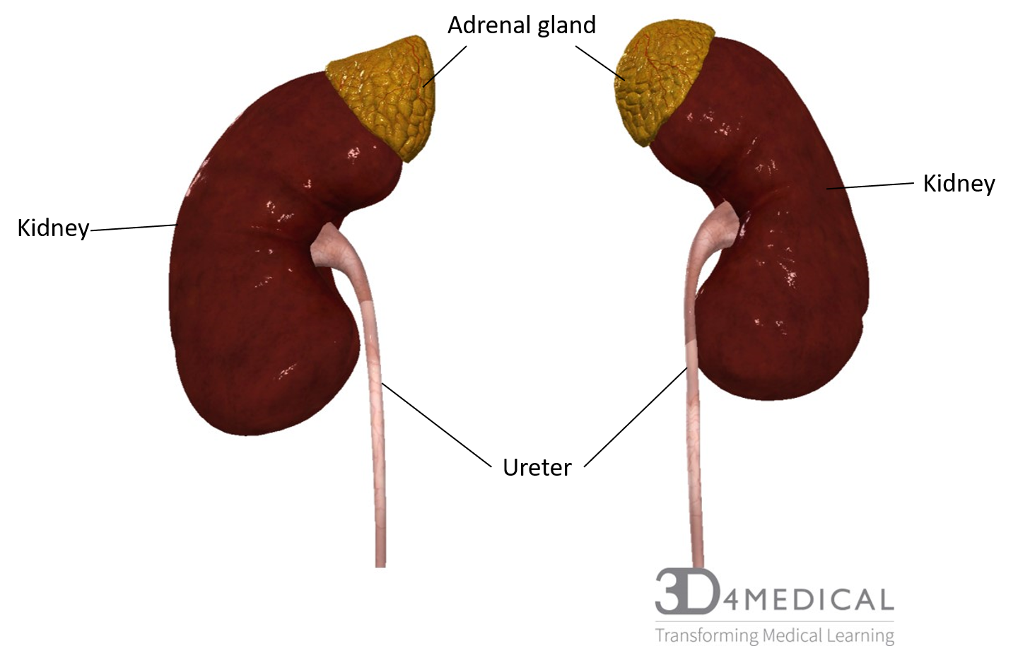
The adrenal gland is a yellow, pyramid-shaped gland that sits on top of each kidney. Since they sit on top of the kidney’s superior border, the adrenals can be referred to as the suprarenal gland. The adrenal glands are connected to the kidneys via the dense fibrous capsule that firmly holds the two organs together. The adrenals are also located near the 12th rib and the diaphragm with major arteries and veins in close proximity as they run along the posterior wall of the abdominopelvic cavity. Since it is an endocrine organ it is important that is highly vascularized, this will allow for the secreted hormones to affect the body. The adrenal gland had two key layers, the adrenal medulla and adrenal cortex.
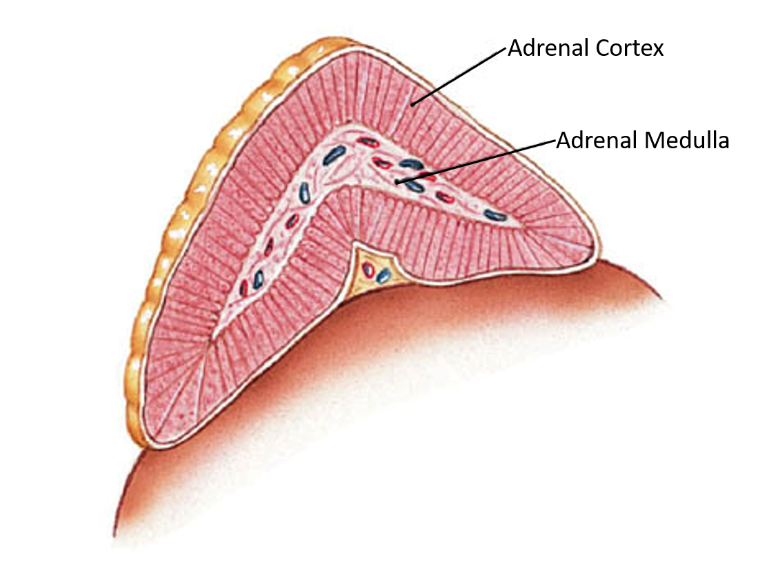
The adrenal medulla is the deep layer of the adrenal gland. The medulla is often a grey-pink colour due to the abundance of blood vessels and connective tissues. The medulla is responsible for the secretion of two hormones. From specialized cells epinephrine, also known as adrenaline, and norepinephrine, also known as noradrenaline, are produced. Each hormone is produced by one cell with evidence showing that the two types of cells are found in different areas of the adrenal medulla and that they are independently controlled.
The adrenal cortex is the more superficial layer. It has a yellowish colour due to the stored lipids, which include cholesterol and various fatty acids. From the cortex, dozens of hormones are released that are collectively called corticosteroids. Corticosteroids are vital to survival. These corticosteroids are produced in three distinct zones on the adrenal cortex, these include the zona glomerulosa, zona fasciculata, and zona reticularis. Each zone produces specific steroid hormones that make up the corticosteroids. Transport proteins are needed to move adrenal hormones around the body. Transcortin, found in the blood bound with the corticosteroid hormones and carry them to their target organs.
Hormones
The adrenals medulla is responsible for the release of epinephrine and norepinephrine. These hormones are released by packing the hormones into vesicles that then cluster just inside the plasma membrane. From here these hormones are slowly released at low levels through exocytosis, however, if there is a sympathetic stimulus epinephrine and norepinephrine are released at a dramatically accelerated rate of exocytosis. The sympathetic nervous system involves the fight or flight mechanism, so it makes sense that the role of epinephrine and norepinephrine help the body response to a stressful situation. Epinephrine and norepinephrine have four key functions, the acceleration of ATP production, breakdown of stored fat, liberating glucose stores in the liver, and increase the rate and force of cardiac muscle contraction. This in terms of short-term stress this response means that your heart rate and pressure will increase and that you will have better muscular strength and endurance. So, if your stressful situation is running into a hungry lion or mugger you will have the surge of strength and endurance to fight your attacker or run away. The metabolic changes that epinephrine and norepinephrine cause reach their peak 30 seconds after the stimulus and persist for several minutes.
The adrenal cortex has a more complex role when it comes to the endocrine functions. It is responsible to produce corticosteroids which can be broken down into three groups that are associated with the zone that produces them. These zones are the Zona Glomerulosa, Zona Fasciculata and the Zona Reticularis.
The Zona Glomerulosa is responsible for the production of a group of hormones called Mineralocorticoids. The mineralocorticoids affect the concentration and composition of electrolytes in body fluids. The main hormone from mineralocorticoid group is Aldosterone. Aldosterone is responsible for the conservation of sodium ion and the elimination of potassium ion. The target of aldosterone is the kidneys, sweat gland, salivary gland and the pancreas. Aldosterone is also responsible for increasing a person interest in salty food by increasing the sensitivity of the salt receptor taste bud on the tongue. Aldosterone is stimulated when the sodium concentration falls, a drop in blood volume, a drop in blood pressure, or a rise in potassium concentration in the blood.
The Zona Fasciculata is responsible for the production of the group of hormones called Glucocorticoids. Glucocorticoids are a group of steroid hormones that affect glucose metabolism. When the zona fasciculata is stimulated by ACTH, a pituitary hormone, the secretion of cortisol also known as hydrocortisone. Cortisol is the primary hormone in the glucocorticoid, with corticosterone and cortisone also released in smaller amounts. Cortisol is often referred to as the stress hormone, but unlike epinephrine and norepinephrine, the stressor is not a single short-term event. In times of long-term stress, cortisol helps control blood sugar levels, regulate metabolism, reduce inflammation and assist in memory formulation.
The final zone is Zona Reticularis, this zone produces small amounts androgens. Androgens are usually associated with the reproductive organs, but small amounts are produced by the adrenal cortex. In early stages of puberty adrenal androgen stimulate the growth of pubic hair in both girls and boys. In adulthood, adrenal androgens are not important to men. However, for adult women, adrenal androgens are important to build muscle mass, blood cell formation and to support their sex drive.
Clinical application
In today’s world chronic stress is commonplace, with people stressing about finances, school, work, family and ironically health. Although cortisol can be helpful in short bouts of chronic stress, it can be damaging when high levels are maintained over a long period of time. In this short video by DNews explains the effect of long-term stress.
References
https://www.hormone.org/hormones-and-health/hormones/cortisol
Bartholomew E.F., Hutching R.T. Martini F.H., Nath J.L, Ober C.E.,Ober W.O, O’Keefe R.A,& Welch K.. (2015) Fundamentals Of Anatomy and physiology (10th ed.) Pearson
Videos:
D News https://www.youtube.com/watch?v=8IUZjCSkbrc
Pancreas
Organs
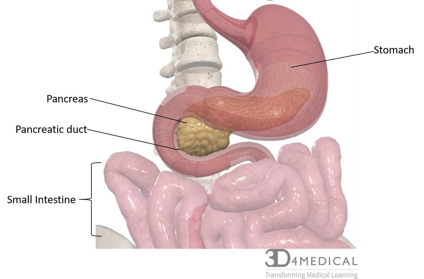
The pancreas is both an exocrine digestive organ and endocrine, playing a major role in internal metabolism and external digestion. The pancreas is part of the retoparitanemum as it sits deep to the stomach and proximal portion of the small intestine called the duodenum. The pancreas is long, slender, lumpy and pale organ ranging from 20-25 cm long with an average weight (in adults) of 80g. There are three sections to the pancreas, the head, the body and the tail. The head fits into the loop formed by the duodenum. The body it the middle section that extends toward the spleen, this section forms the long slender shaper of the pancreas. Lastly, the tail is the most lateral section of the pancreas and forms a short, blunt, rounded end of the pancreas.
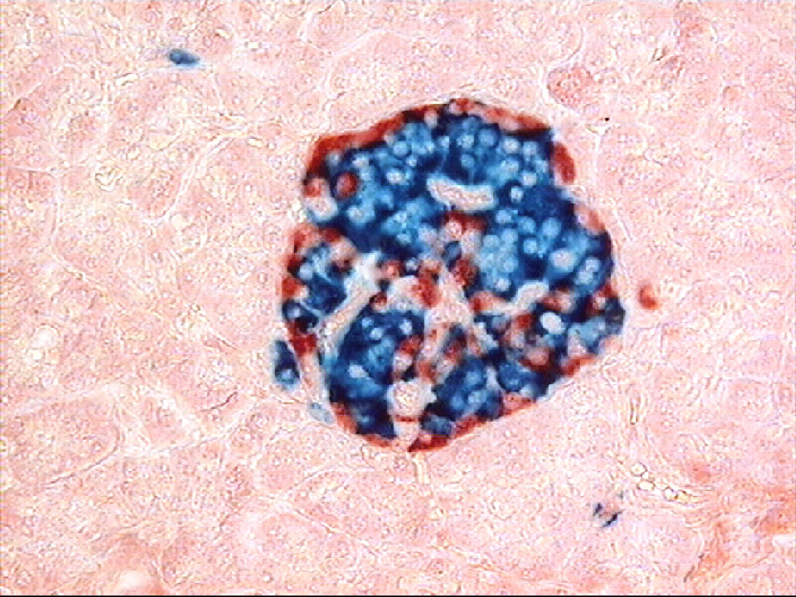
The endocrine functions of the pancreas are based on a cluster of cells called the pancreatic islets or the islets of Langerhans. Although only about 1% of the pancreas is made up of the islet of Langerhans, a typical pancreas can contain 2 million islets of Langerhans. They are four different types of cells in the islet of Langerhans, these cells are called Alpha cells, Beta cells, Delta cells and F cells. Alpha cells and beta cells are responsible for regulating the blood sugar levels. Delta cells produce peptide hormone identical to growth hormone- inhibiting hormone (GH-IH) which suppresses the release of glucagon and insulin. A peptide hormone released from the delta cells also slow the rated of food adsorption and enzyme secretion along the digestive tract. The second cell type found in the Pancreatic acini is the F cells. The F cells produce the hormone pancreatic polypeptide (PP) which inhibits gallbladder release of bile and regulates the production of some of the pancreatic enzymes. This is thought to help to control the rate of nutrient absorption.
The pancreas also has a role in exocrine digestion through the pancreatic acini clusters can be found around the ends of the pancreatic ducts. The pancreas plays an important in digestion as it produces digestive enzymes and buffers. For more information about the exocrine function of the pancreas go to the visceral organ section under the Abdominal tab.
As the pancreas is an endocrine gland a good blood supply is necessary to move the hormones produced by the pancreatic islets to the rest of the body. The arteries that feed the pancreas are the splenic, superior mesenteric, and common hepatic arteries. These arteries branch off to form the major branches called the pancreatic arteries and the pancreaticoduodenal arteries. The venous system removes wastes and moves hormones around the body. The splenic view and its branches move waste and hormones away from the pancreas to be excreted or used.
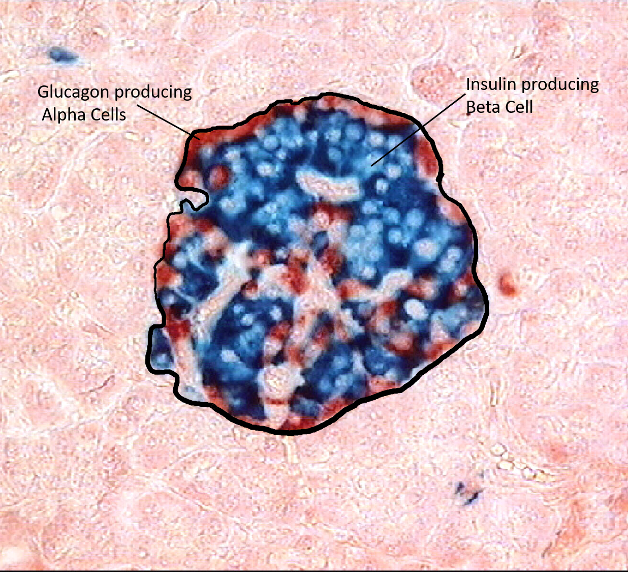
Image of an islet of Langerhans that is died to show where the hormones are found.
Hormones
The pancreas is responsible for hormones that are responsible for the metabolism of the glucose in the blood. The two main hormones are insulin and glucagon. They work together to regulate blood sugar levels. These hormones are produced in the islets of Langerhans by specialized cells. The beta and alpha cells. Together these two hormones work to maintain a normal blood sugar level ranging between 70-110 mg/dL of blood. This is done through be each hormone’s effect on lean tissues, adipose tissue, and liver.
Insulin is a peptide hormone that is responsible for the reduction of high blood sugar, secreted by the beta cells. To drop the blood sugar level in the blood, insulin is released to increase the utilization of glucose by the lean tissues and through the storage of excess glucose. Insulin can be thought of as a key that unlocks the cells allowing glucose into the cells. Glucose is necessary for energy production so lean tissues like muscles, especially the heart muscles, use a lot of glucose. In the muscle and liver excess glucose is stored as glycogen. This storage is an important immediate source of energy in times of stress on the muscles, like physical activity. If there is excess glucose left in the blood that hasn’t been used by the lean tissues or stored in the muscle or liver, insulin is also responsible for the storing the excess glucose as fat. The adipose tissues are very sensitive to insulin. Insulin has 6 effects the target cells that are dependent on the cell type of the target. These include acceleration of glucose uptake, accelerating glucose utilization, enhanced ATP production, stimulating glycogen formation, stimulating amino acid absorption and protein synthesis, and stimulating triglyceride formation in adipose tissue. Problems arise when insulin is unable to get sugar out of the bloodstream. See the clinical application for an explanation on how the body responses to high blood sugar.
Although high blood sugar can to problematic, low blood sugar can also impact our health. To prevent low blood sugar and to maintain homeostasis within the normal blood sugar range, glucagon is released by the alpha cells. A way to remember the stimulus for the release of glucagon is to break the word do into to an acronym. Glucagon is released when Glucose is All Gone. The primary function of glucagon is to increase blood sugar and to do that is has 3 key effects: to stimulate the breakdown of glycogen, stimulating the breakdown of triglycerides in adipose tissue, and stimulate the production and release of glucose by the liver. The process that glucagon stimulates to increase blood sugar levels is called gluconeogenesis. Gluconeogenesis is the process that happens in the liver to reform glucose from its storage form, especially when it is stored with amino acids.
Clinical application
Diabetes Mellitus commonly referred to as diabetes, sometimes referred to as the eye, nerve and kidney disease, is caused by the inability to lower the blood sugar levels. There are two types of Diabetes mellitus, type I and type 2.
Type 1, which used to be referred to as adolescent onset diabetes, is when beta cells don’t produce adequate amounts of insulin. Often this shows up in childhood or as a young adult and requires lifelong blood sugar management through insulin injections and dietary changes. Type 1 diabetes accounts for approximately 5% of cases.
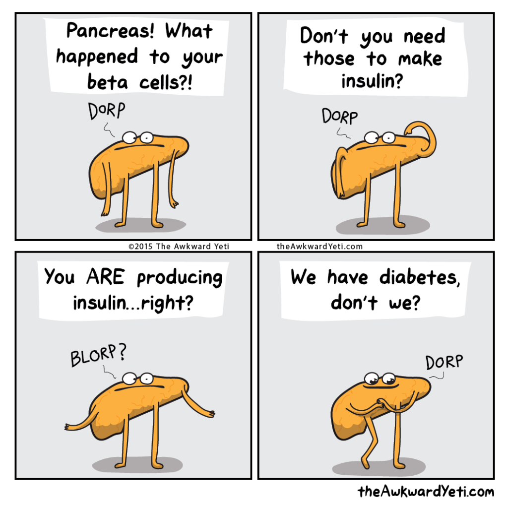
Type 2 diabetes, or adult-onset diabetes, is considered a lifestyle caused disease as type 2 diabetes is associated with obesity. Type 2 Diabetes is caused by the insulin targets cells becoming insensitive to the stimulus, this, in turn, means that glucose remains in the blood causing elevated blood sugar. The elevated blood sugar can cause damage to nerves in the eyes, peripheral nerves, heart, peripheral tissue damage and kidney damage. The damage to the peripheral nerves and tissues often are associated (incomplete sentence). In people with type 2 diabetes injury to the feet can go unnoticed due to the loss of nerve function. Injuries even if treated can take a long time to heal as the elevated blood sugar slows the healing process. However, unlike type 1 diabetes, type 2 is often curable. Through weight loss associated with healthy lifestyle changes type 2 diabetes can be reversed.
Type 2 diabetes, or adult-onset diabetes, is considered a lifestyle caused disease as type 2 diabetes is associated with obesity. Type 2 Diabetes is caused by the insulin targets cells becoming insensitive to the stimulus, this, in turn, means that glucose remains in the blood causing elevated blood sugar. The elevated blood sugar can cause damage to nerves in the eyes, peripheral nerves, heart, peripheral tissue damage and kidney damage. The damage to the peripheral nerves and tissues often are associated (incomplete sentence). In people with type 2 diabetes injury to the feet can go unnoticed due to the loss of nerve function. Injuries even if treated can take a long time to heal as the elevated blood sugar slows the healing process. However, unlike type 1 diabetes, type 2 is often curable. Through weight loss associated with healthy lifestyle changes type 2 diabetes can be reversed.
References
Bartholomew E.F., Hutching R.T. Martini F.H., Nath J.L, Ober C.E.,Ober W.O, O’Keefe R.A,& Welch K.. (2015) Fundamentals Of Anatomy and physiology (10th ed.) Pearson
Bartholomew E.F., Hutching R.T. Martini F.H., Nath J.L, Ober C.E.,Ober W.O, O’Keefe R.A,& Welch K.. (2015) Fundamentals Of Anatomy and physiology (10th ed.) Pearson
https://www.hormone.org/hormones-and-health/hormones/insulin
needs citation https://nyaspubs.onlinelibrary.wiley.com/doi/abs/10.1111/j.1749-6632.1959.tb44921.x
video:
khan academy https://www.youtube.com/watch?v=xNf–q0YMq8
Pictures:
Picture of the islet of Langerhans https://upload.wikimedia.org/wikipedia/commons/0/05/Human_pancreatic_islet.jpg
