Upper Limb
Muscles
As we have mentioned in previous sections, the pectoral girdle or the shoulder girdle sacrifices structural integrity for a greater range of motion. Despite this being the case the pectoral girdle maintains a large degree of stability mostly due to intricate musculature of the region, all while allowing for some unique movement. This was an evolutionary trait as a result of the upper limbs no longer needing to be weight bearing limbs; now instead of walking using our arms, we became able to hunt, gather and undertake difficult tasks more efficiently with our upper limbs. These muscles can be divided up into two groups based on their actions, muscles that position the girdle, more specifically the clavicle and the scapula, and muscles that move the arm.
Muscles Positioning The Shoulder
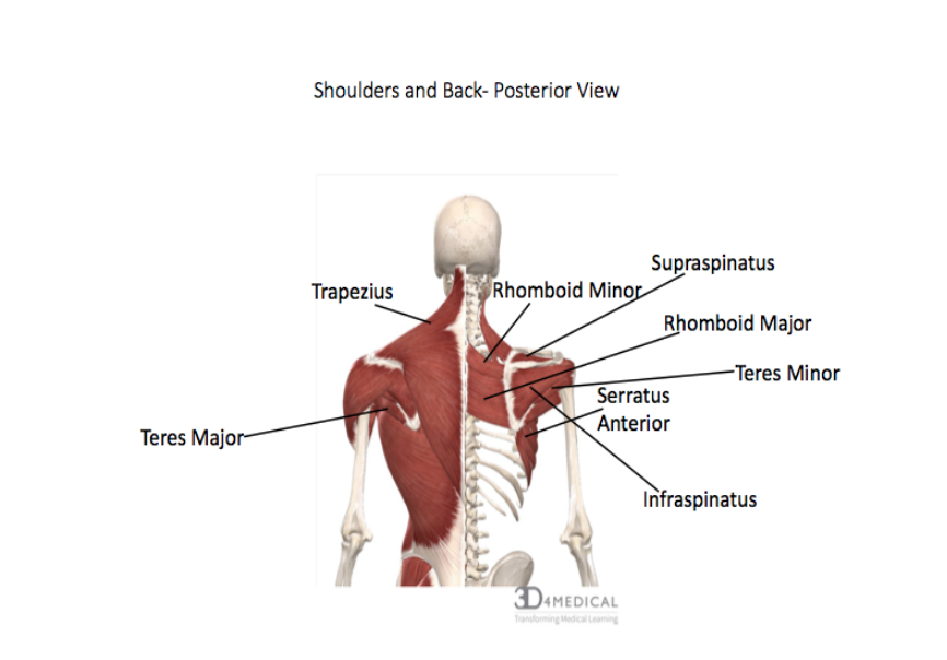
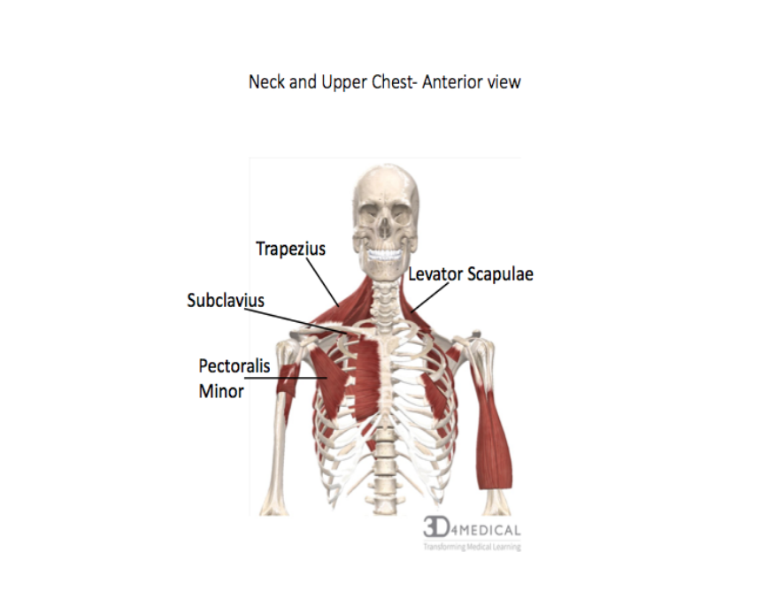
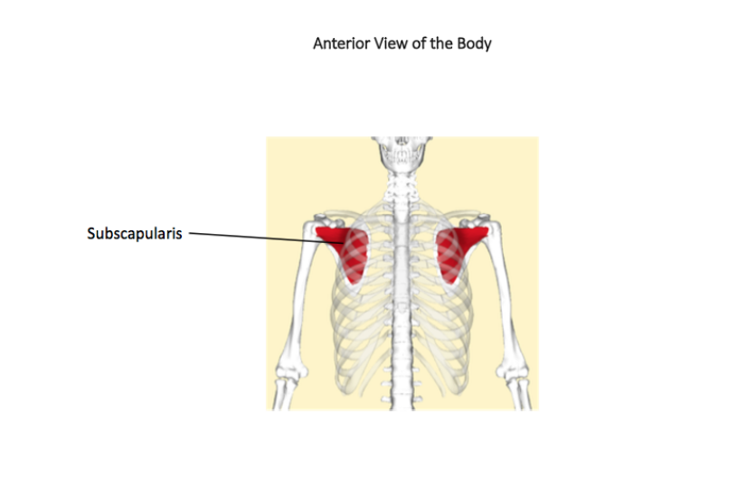
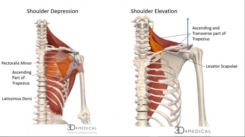
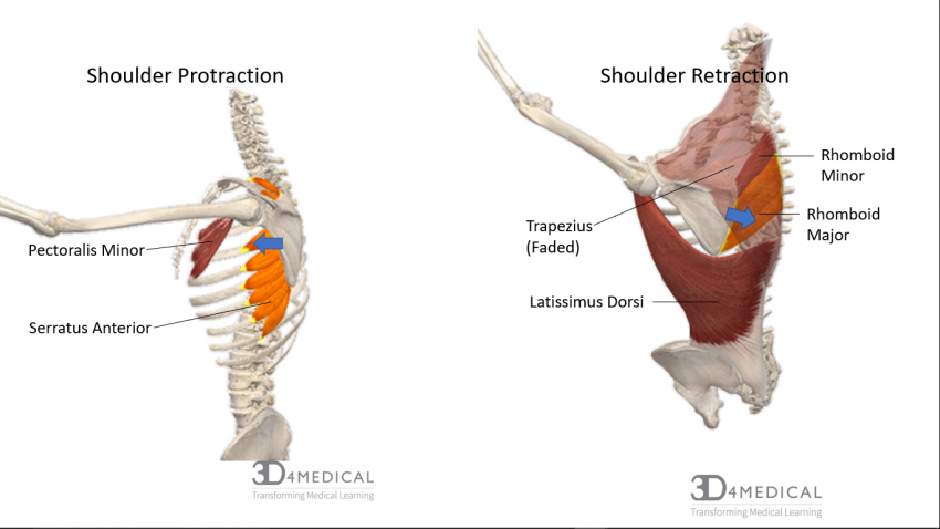
Muscles that position the pectoral girdle are all stated in great detail in the first table below, we will quickly explore some of them, their unique aspects, and their actions. Beginning with one of the most dominant muscles in the region, the trapezius. This is a large, triangular in shape, and flat muscle; its actions can vary based on which region of the muscle is active. This means that the trapezius muscle can be divided into 3 portions, the ascending, transverse, and descending parts, also known as the inferior, middle, and superior parts respectively. This muscle stabilizes and secures the scapula on the thoracic cage, scapular protraction, and retraction, as well as elevation and depression. Next, we have rhomboids major and minor. The rhomboids are two diamond-shaped muscles that extend from the vertebral column to the medial border of the scapula; generally, there appears to be a gap between them but sometimes this is not present and they look like one large muscle. Both muscles, as evident by their orientation, cause retraction of the scapula, as well as its rotation when lowering the arm from a raised position. A lot like the trapezius, the rhomboids can also stabilize the scapula on the rib cage. Another shoulder positioning muscle that can be observed on the posterior portion of the pectoral girdle is levator scapulae. As the Latin origin word “levator” suggests the main action of this muscle is to elevate the scapula. This is clear when observing its attachment site on the superior border of the scapula. The levator scapulae also compliments the rhomboids in their action of rotating the shoulder blade when lower the arm from an elevated position. A somewhat “hidden” muscle in this group is the subscapularis muscle, which attaches on the anterior aspect of the scapula, sandwiching it between the shoulder blade and the rib cage. It stabilizes the shoulder, but also assists in arm movements that we will explore shortly. Moving to the anterior portion of the “muscles that position the shoulder” grouping in the pectoral girdle we can observe the pectoralis minor muscle. This muscle is inferior to the famous pectoralis major which we will also discuss shortly. Pectoralis minor is a “fan” shaped muscle that assists in pulling the scapula anteriorly or “protracting” it, as well as depression of the shoulder. Laying behind pectoralis minor is the small subclavius muscle. This muscle primarily supports the clavicle at the sternoclavicular joint during movements of the upper limb. The final muscle we will talk about in this group is the serratus anterior, perhaps the most interesting looking muscle in the body. The edge of this muscle can be resemblant of a serrated knife. It is dubbed the “boxer muscle” because it is largely responsible for scapular protraction when throwing a punch; we do not endorse you attempt this with another individual to test this out. This action can be explained by the muscle’s attachment on the anterior aspect of the medial border of the scapula and wrapping around the outside of the rib cage.
Muscles That Move The Arm
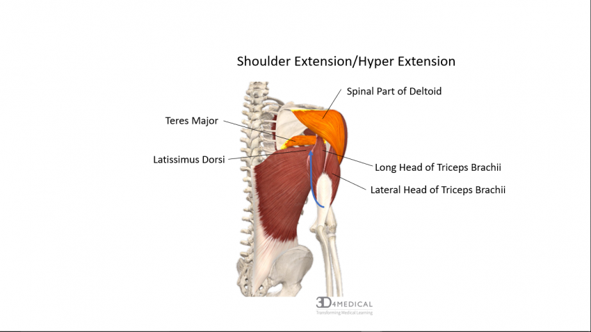
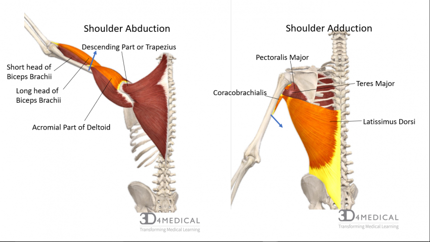
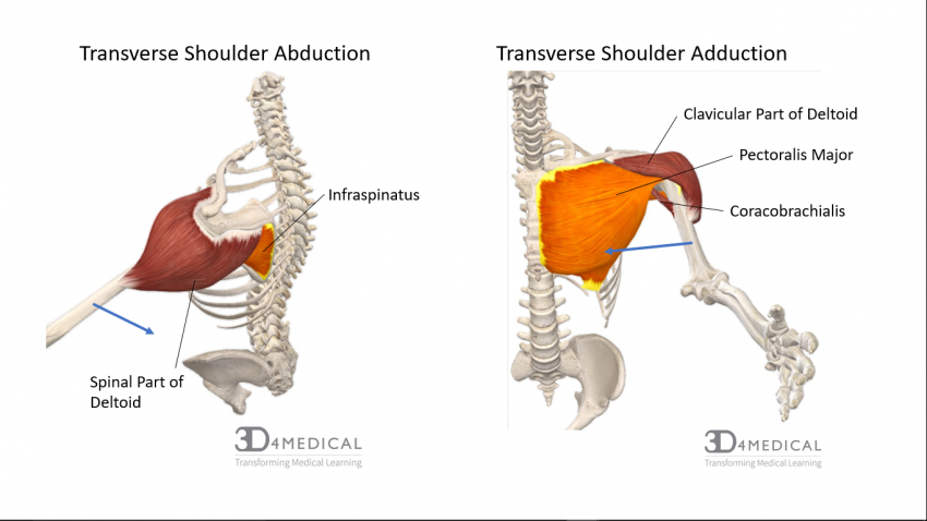
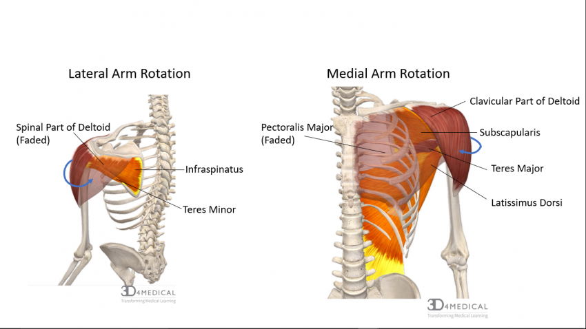
The second grouping of muscles in the pectoral girdle are the muscles that assist in arm movement. A commonly known group within these muscles is the “rotator cuff”. The rotator cuff consists of the supraspinatus, infraspinatus, teres minor, and subscapularis muscles; this grouping is also often abbreviated as “SITS” standing for the first letter in each muscle’s name. The supraspinatus, as the name implies, sits above the spine of the scapula in the supraspinous fossa, and goes around the outside of the humerus and inserts at the greater tuberosity of the humerus, along with infraspinatus and teres minor. The infraspinatus sits in the infraspinatus fossa, while teres minor sits slightly inferiorly to it. The odd one out in this group is the subscapularis muscle we spoke about earlier in the “muscles that position the shoulder” grouping; it instead inserts on the lesser tuberosity or tubercle. This group works in conjunction to externally rotate the upper arm and they pull the head of the humerus into the glenoid cavity. The next muscles on our list is teres major, which also assists the rotator cuff group in stabilizing the shoulder joint. Its main actions include adduction and internal rotation of the humerus, as well as scapular protraction. Another large muscle in this region is the deltoid muscle. It is located on the upper part of the arm atop the shoulder, and it is named after the Greek letter delta due to its equilateral triangle shape resembling the rounded triangular shape of the deltoid itself. This muscle can flex and medially rotate the arm when its anterior portion is activated, its lateral portion abducts the arm, and its posterior portion can extend and laterally rotate the arm. Finally, we look at the pectoralis major muscle, which can be subdivided into its clavicular head and its sternocostal head. The clavicular portion of pectoralis major flexes the humerus, while the sternocostal head adducts and medially rotates the humerus.

Muscles That Position The Shoulder
| Muscle | Origin | Insertion | Action | Innervation |
| Levator Scapulae | Transverse Process of C1-C4 | Vertebral Border of Scapula Near the Superior Angle | Elevates the Scapula | C 3,4 and Dorsal Scapular Nerve C5 |
| Pectoralis Minor | Anterior Superior
Aspect of Ribs 2 to 5 |
Coracoid Process of Scapula | Depresses the Shoulder,
Rotates Scapula Inferiorly |
Medial Pectoral Nerve
(C8-T1) |
| Rhomboid Major | Spinous Process of Superior Thoracic Vertebra | Vertebral Border of Scapula Below the Spine | Adduction &
Downward Rotation of the Scapula |
Dorsal Scapular Nerve
C5 |
| Rhomboid Minor | Spinous process of C7,T1 | Vertebral border of scapula near the spine | Adduction & downward rotation | Dorsal Scapular nerve C5 |
| Serratus Anterior | Anterior & Superior Margins of Ribs 1- 8 | Anterior Surface of Vertebral Border of Scapula | Protracts Shoulder &
Upward Rotation of Scapula |
Long Thoracic Nerve
T 5-7 |
| Trapezius | Occipital Bone Ligamentum Nuchae & Spinous Process of Thoracic Vertebrae | Clavicle & Scapula
(Acromion and Spine) |
Elevates,
Retracts & Depresses, Rotates the Scapula Upward & Extends the Neck |
Accessory N. (XI) and C 3-4 |
| Subclavius | 1st Rib | Inferior Border of the Clavicle | Depresses & Protracts the Shoulder | Nerve to Subclavius (C5-6) |
| Subscapularis | Subscapular Fossa of the Scapula | Lesser Tubercle of the Humerus | Medial Rotation of the Shoulder | Sub-Scapular Nerve
(C5-6) |
Muscles That Move The Arm
| Muscle | Origin | Insertion | Action | Innervation |
| Deltoid | Clavicle
(Acromion) & Scapular Spine |
Humerus (Deltoid
Tuberosity) |
Abduction,
Flexion & Medial Rotation Extension & Lateral Rotation |
Axillary Nerve (C5-6) |
| Supraspinatus | Supraspinous Fossa of the Scapula | Greater Tubercle of the Humerus | Abduction of the Shoulder | Suprascapular Nerve
C5 |
| Teres Major | Inf. Angle of
Scapula |
Intertubercular Groove of the Humerus | Extension, Adduction & Medial Rotation | Lower Subscap. N.
C 5-6 |
| Infraspinatus | Infraspinous Fossa | Greater Tubercle of the Humerus | Lateral Rotation of the Shoulder | Suprascapular Nerve (C5-6) |
| Teres minor | Lateral border of scapula | Greater tubercle of the humerus | Lateral rotation of the shoulder | Axillary nerve
C5 |
| Pectoralis Major | Ribs 2-6,
Sternum & Medial of the Clavicle |
Greater Tubercle
& Intertubercular Groove |
Flexion, Adduction & Medial Rotation of the Shoulder | Pectoral Nerve
C5-T1 |
These are not the only muscles that undertake such actions on the arm. Later in this section, you will explore the brachial region which will go into more detail about other muscles that move the arm.
Muscles Of the Brachium
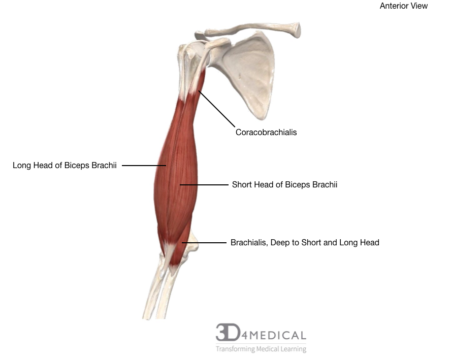
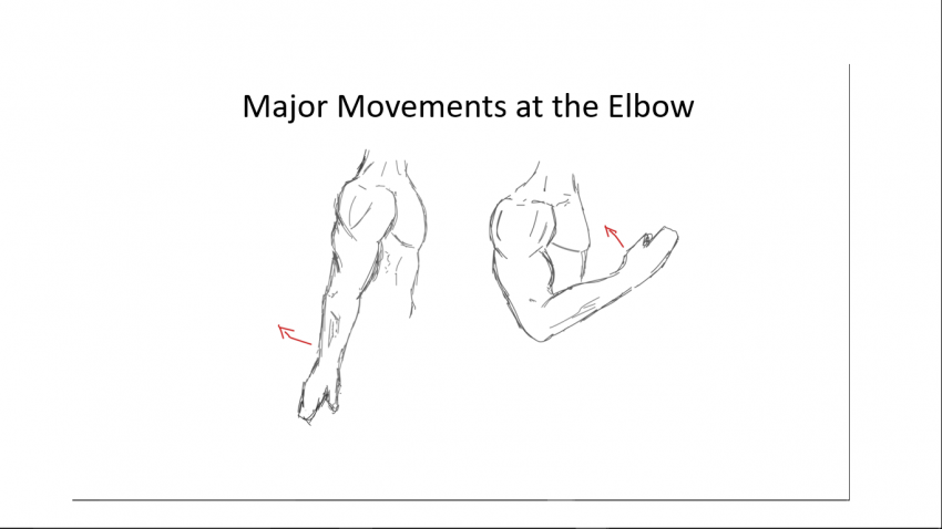
The four muscles of the arm are the biceps brachii, brachialis, coracobrachialis, and triceps brachii. Of the four, the biceps brachii, coracobrachialis, and brachialis are found in the anterior compartment while the triceps brachii can be found in the posterior compartment.
Biceps Brachii
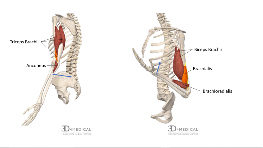
As the name bicep suggests, this muscle contains two muscle bellies (long head, short head). The long head and short head of biceps brachii meet together at the midpoint of the humerus. The primary action of this muscle is flexion at the elbow and shoulder joint and supination of the anti-brachium.
Brachialis
The brachialis sits deep to the biceps brachii muscle. Meaning underneath. The main function of the brachialis is to keep the forearm stable during both slow and rapid movements. During flexion of the elbow, the brachialis always contracts and remains in flexion until the movement is finished. These traits are the reason the brachialis is regarded as one of the hardest working muscles in the arm.
Coracobrachialis
The coracobrachialis is also involved in the flexion and abduction of the arm and also helps to stabilize the shoulder joint.
The image below showcases the anterior compartment of the arm including the three muscles mentioned above.
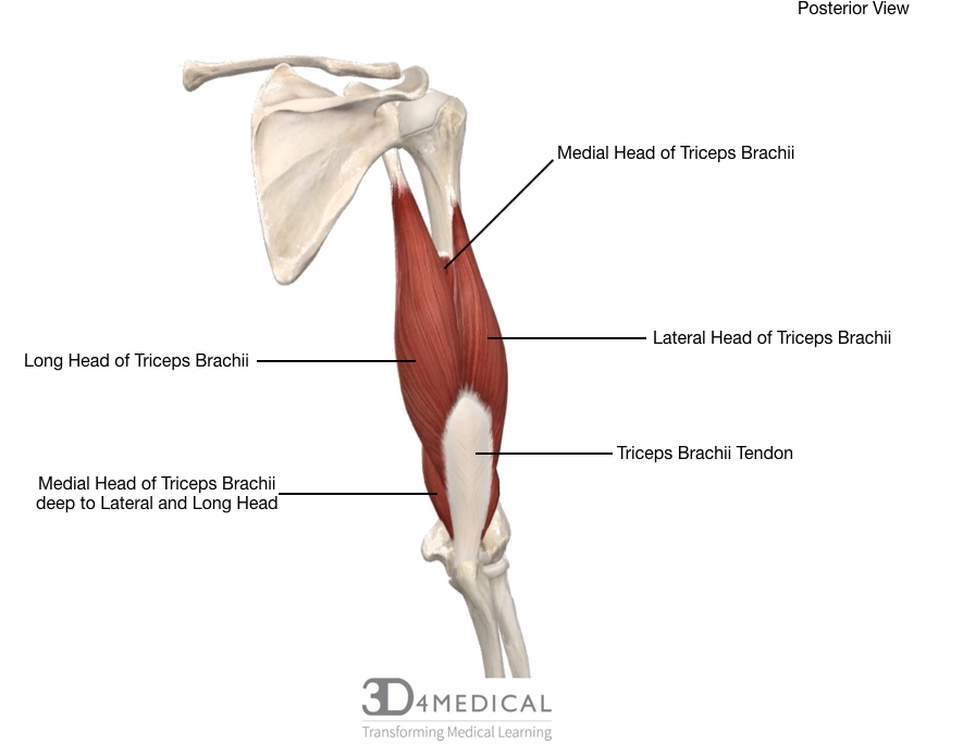
Triceps Brachii
This muscle is the only muscle included in the posterior compartment. Like biceps brachii, triceps also have numerous muscle bellies. Tri meaning three; long head, lateral head, and a medial head which lies deep to the lateral and long head. This muscle is the main extensor at the elbow joint and is the antagonist to the bicep brachii meaning it counteracts its movement. The triceps also help with extension and adduction of the arm.
Anconeus
This muscle is a very small muscle that is located on the posterior portion of the elbow. Some refer to this muscle as a continuation of the triceps muscle. It aids in the extension of the forearm, stabilizes the elbow joint and also abducts the ulna in pronation.
The image below showcases the triceps brachii in the posterior compartment.
| Muscles of the Brachium | ||||
| Muscle | Origin | Insertion | Action | Innervation |
| Biceps Brachii | Long Head: Supraglenoid Tubercle
Short Head: Coracoid Process |
Radial Tuberosity and Bicipital Aponeurosis | Shoulder Flexion, Elbow Flexion and Supination | Musculocutaneous Nerve |
| Brachialis | Anterior Surface of Distal Half of Humerus | Coronoid Process of Ulna | Flexion at Elbow Joint to Flex Forearm | Musculocutaneous Nerve |
| Coracobrachialis | Coracoid Process of Scapula | Middle Third of Medial Surface of Humerus | Flexion, Adduction, Internal Rotation | Musculocutaneous Nerve |
|
Triceps Brachii |
Lateral Head:
Posterior Surface of Humerus that Lies Superior to Radial Groove Long Head: Infraglenoid Tubercle Medial Head: Posterior Part of Humerus that Lies Inferior to Radial Groove |
Olecranon process of ulna via triceps brachii tendon | Lateral Head: Extension at Elbow Joint to Extend Forearm
Long Head: Extension and Adduction of Shoulder Joint, Extension at Elbow Medial Head: Extension at Elbow Joint to Extend Forearm |
Radial Nerve |
| Anconeus | Lateral Epicondyle of Humerus | Lateral Surface of Olecranon; Posterior Surface of Superior Part of Ulna | Extension of Forearm | Radial Nerve |
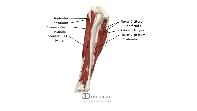
Muscles of the Forearm
The muscles that originate in this area are responsible for flexion and extension of the wrist. There are also muscles for pronation and supination of the hand, as well as movements of the hand.
| Muscle | Origin | Insertion | Action | Innervation |
| Flexor Carpi Radials | Medial Epicondyle of the Humerus | Base of Second and Third Metacarpals | Flexes and Abducts Wrist | Median Nerve |
| Flexor Carpi Ulnaris | Medial Epicondyle of the Humerus and Olecranon Process | Carpal and Metacarpal Bones | Flexes and Adducts the Wrist | Ulnar Nerve |
| Palmaris Longus
|
Medial Epicondyle of the Humerus | Fascia of Palm | Flexes Wrist | Median Nerve |
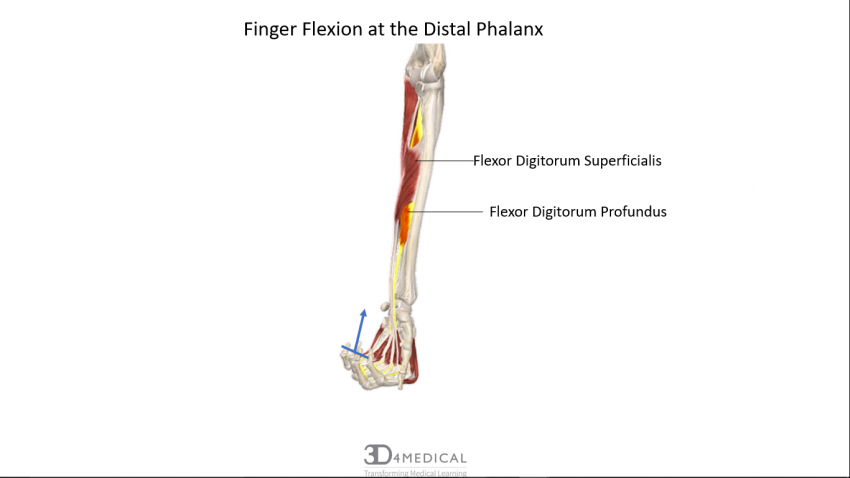
| Flexor Digitorum Profundus | Anterior Surface of Ulna | Bases of Distal Phalanges in Fingers two through five | Flexes Distal Joint of Fingers | Median, Ulnar Nerve |
| Flexor Digitorum
Superficialis |
Medial Epicondyle of the Humerus, Coronoid Process of Ulna, and Radius | Tendons of Finger | Flexes Fingers and Hand | Median Nerve |
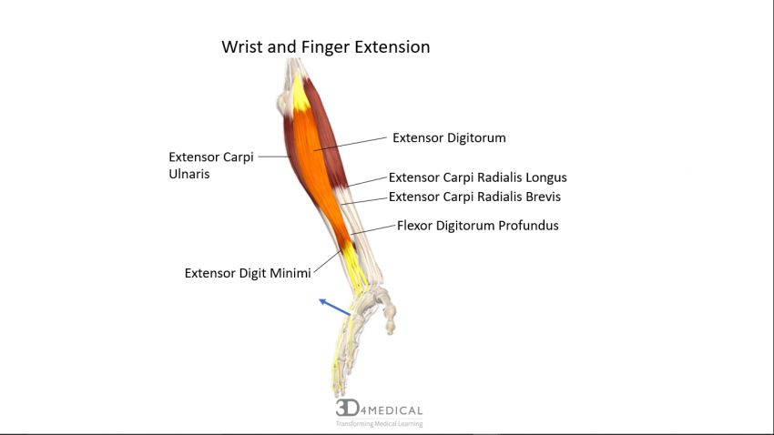
| Extensor Carpi Radialis Longus | Distal End of Humerus | Base of Second Metacarpal | Extends Wrist and Abducts Hand | Radial Nerve |
| Extensor Carpi Radialis Brevis | Lateral Epicondyle of Humerus | Base of Second and Third Metacarpals | Extends Wrist and Abducts Hand | Radial Nerve |
| Extensor Carpi Ulnaris | Lateral Epicondyle of Humerus | Base of Fifth Metacarpal | Extends and Adducts Wrist | Radial Nerve |
| Extesnor Digitorum | Lateral Epicondyle of Humerus | Posterior Surface of Phalanges in Fingers Two Through Five | Extends Fingers | Radial Nerve |
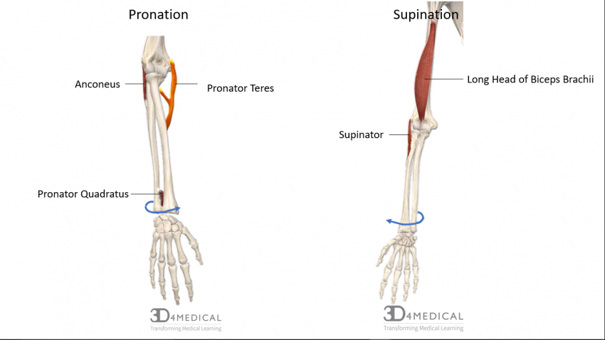
| Pronator Quadratus | Anterior and Medial Surfaces of Distal Portion of Ulna | Anterolateral Surface of Distal Portion of Radius | Pronation | Median Nerve |
| Pronator Teres | Medial Epicondyle of Humerus and Coronoid Process of Ulna | Midlateral Surface of the Radius | Pronation | Median Nerve |
| Supinator | Lateral Epicondyle of Humerus, Annular Ligament, and Ridge Near Radial Notch of Ulna | Anterolateral Surface of Radius Distal to the Radial Tuberosity | Supination | Deep Radial Nerve |

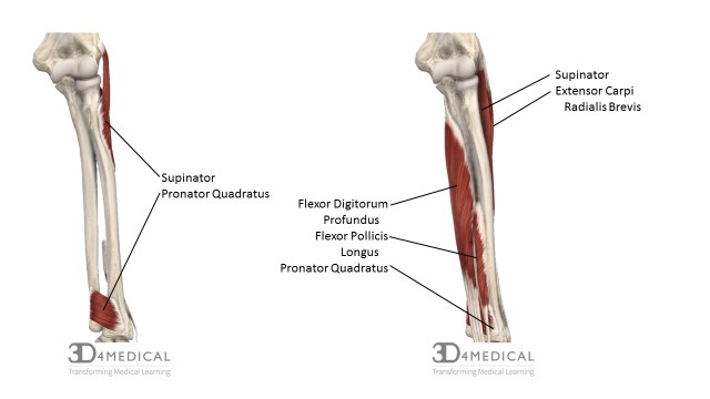
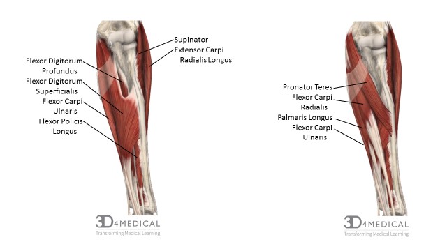

(Muscles of the anterior forearm beginning deep (top) and moving superficial (bottom).)
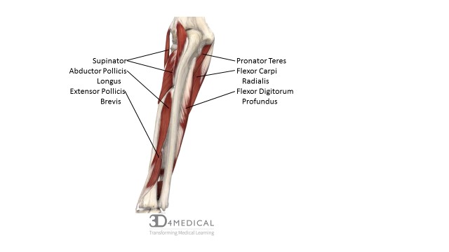
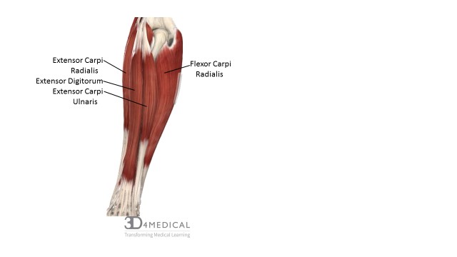
(Muscles of the posterior forearm beginning deep (top) and moving distal (bottom).)
Extrinsic Muscles of the Hand
The muscles that reside within this area are varied and unique by which they act on the digits from an origin outside of the hand and insert within it. The groups of muscles within this area are therefore referred to as the extrinsic muscles of the hand. Based on their respective locations and functions, the muscles of the forearm are thereby divided into two compartments: 1) anterior compartment muscles, and 2) posterior compartment muscles. For further insight regarding the muscles of the forearm check out Jason’s section.
Intrinsic Muscles of the Hand
As discussed in the prior section regarding the muscles of the forearm, granted they produce far more powerful movement, but in turn they sacrifice a deftness of movement that only the intrinsic muscles of the hand can produce. Thusly, the intrinsic muscles of the hand are divided into three compartments: 1) hypothenar, 2) intermediate, and 3) thenar.
The hypothenar aspect of the palm includes: abductor digiti minimi, flexor digiti minimi brevis, and opponens digiti minimi. This is the medial rounded contour of the palm, otherwise termed as the “ball of the little finger.”
| Hypothenar (Medial Aspect of Palm) | ||||||
| Muscles | Origin | Insertion | Action | Innervation | ||
| Abductor Digiti Minimi | Tendon of Flexor Carpi Ulnaris and Pisiform. | Medial, Proximal Phalanx of ‘Pinky’ Finger. | Abduct and Flex Pinky at Metacarpophalangeal Joint. | Ulnar Nerve. | ||
| Flexor Digiti Minimi Brevis | Hamate and Flexor Retinaculum. | Medial, Proximal Phalanx of ‘Pinky’ Finger. | Flexes Pinky at Carpometacarpal and Metacarpophalangeal Joint. | Ulnar Nerve. | ||
| Opponens Digiti Minimi | Hamate and Flexor Retinaculum. | Medial Aspect of Fifth Metacarpal. | Moves Pinky into Opposition at Carpometacarpal Joint. | Ulnar Nerve. | ||
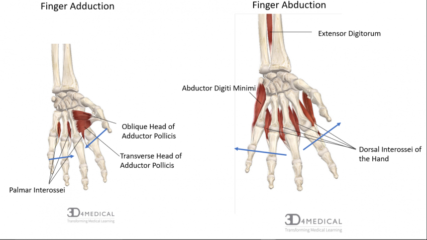
The eleven intermediate muscles of the palm are comprised of the: lumbricals, palmar interossei, and dorsal interossei. Lumbricals originate from and insert into the tendons of other muscles – such as extensor digitorum and flexor digitorum profundus – and are named thusly for their ‘worm-like’ shape. Both sets of interossei muscles are the most posterior groups of the palm; and, their corresponding structure and function allow us to perform such skillful activities such as playing the piano, typing, and writing.
| Intermediate (Midpalmar) | |||||
| Muscles | Origin | Insertion | Action | Innervation | |
| Lumbircals | Lateral Sides of Flexor Digitorum Profundus and Tendons of Each Finger. | Lateral Sides of Extensor Digitorum Tendons on Proximal Phalanges of Each Finger. | Flex Fingers at Metacarpophalangeal Joint; Extend at Interphalangeal Joints. | Median and Ulnar Nerve. | |
| Palmar Interossi | Side shafts of metacarpals of all digits (except middle). | Side Bases of all Proximal Phalanges of all Digits. | Extend Fingers at Interphalangeal Joints; Adduct Each Finger Except the Middle. | Ulnar Nerve. | |
| Dorsal Interossi | Adjacent Sides of Metacarpals. | Proximal Phalanx of Fingers. | Abduct and Flex Fingers 2 – 4 at Metacarpophalangeal Joints; and, Extend Each Finger at Interphalangeal Joints. | Ulnar Nerve. | |
The thenar group of muscles include: abductor pollicis brevis, opponens pollicis, flexor pollicis brevis, and adductor pollicis. The three thenar muscles, as well as the adductor pollicis, come together to create the thenar eminence – or rather, the “ball of the thumb.” Interestingly enough, the adductor pollicis has a fan-like shape and has two heads (transverse and oblique) separated by a gap which allows the radial artery to pass through.
| Thenar (Lateral Aspect of Palm) | ||||||
| Muscles | Origin | Insertion | Action | Innervation | ||
| Abductor Pollicis Brevis | Scaphoid, Trapezium, and Flexor Retinaculum. | Lateral, Proximal Phalanx of Thumb. | Abducts Thumb at Carpometacarpal Joint. | Median Nerve. | ||
| Opponens Pollicis | Trapezium and Flexor Retinaculum. | Lateral Side of Metacarpal I. | Moves Thumb Across Palm in Opposition at Carpometacarpal Joint. | Median Nerve. | ||
| Flexor Pollicis Brevis | Trapezium, Capitate, Trapezoid, and Flexor Retinaculum. | Lateral, Proximal Phalanx of Thumb. | Flexes Thumb at Metacarpophalangeal and Carpometacarpal Joint. | Median and Ulnar Nerves. | ||
| Adductor Pollicis | Oblique Head: Capitate, and Second and Third Metacarpals. Transverse Head: Third Metacarpal. |
Medial, Proximal Phalanx of Thumb via Tendon Containing Sesamoid Bone. | Adducts Thumb at Metacarpophalangeal and Carpometacarpal Joint. | Ulnar Nerve. | ||
https://www.youtube.com/watch?v=WfXTehI1PWo
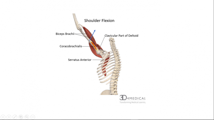
Once shoulder flexion has reached a certain angle, Serratus Anterior contracts to tilt the scapula upwards, allowing for more flexion to occur. If you retract your shoulder (relieving Serratus Anterior) and try to flex it at the same you’ll notice that your arm won’t raise nearly as far.
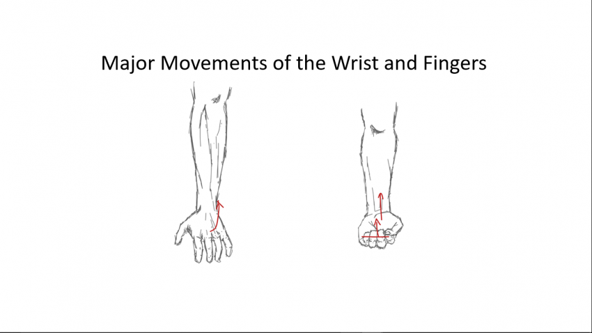
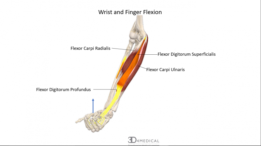
At full contraction these muscles would result in both wrist flexion and finger flexion (such is the case above). However, the wrist or finger can flex independently using the same muscle in a partial contraction, and if antagonist muscles are preventing one movement of the other. The same can be said about extension (see below).