Upper Limb
Surface Anatomy
The pectoral girdle or shoulder girdle is formed by the clavicles, scapulae, and the proximal end of the humerus. This boney structure is surrounded by large muscles that support the girdle. Many of the features of the bones and musculature discussed in earlier sections are palpable, and the media provided will give you a walk through guide. We want to be able to palpate these structures in anatomy because much like looking at a diagram or image, this gives us a point of reference; this makes it easier to learn the anatomy of the region. This allows students to be more aware and involved in their studies, but also it is a tool commonly used by practitioners when working with their patients. So with the permission of a partner walk yourself through some of the steps provided by specialists in the videos, and explore the palpable features of the pectoral girdle. Perhaps when you’ve gotten enough practice with the guides, attempt a walk through on your own and see how many features you can locate.
Were you able to palpate some of the following features?
- Sternoclavicular joint
- Line of clavicle
- Acromial end of the acromioclavicular joint
- Midclavicular line
- Coracoid process – 1 cm medial to glenohumeral joint space
- Greater and lesser tubercles of the humerus
- Scapular spine – medial border – inferior angle – lateral border
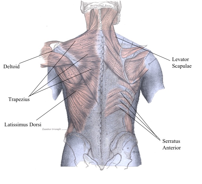
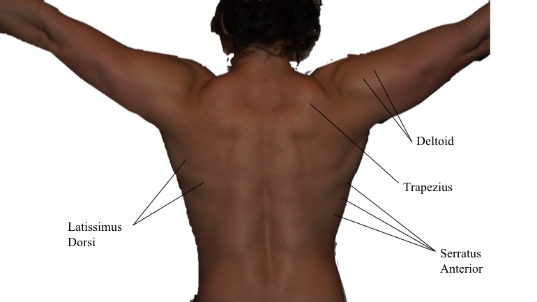
The superficial muscles of the posterior pectoral girdle that can be identified are, Levator Scapulae, Serratus Anterior, Latissmus Dorsi, and Deltoid
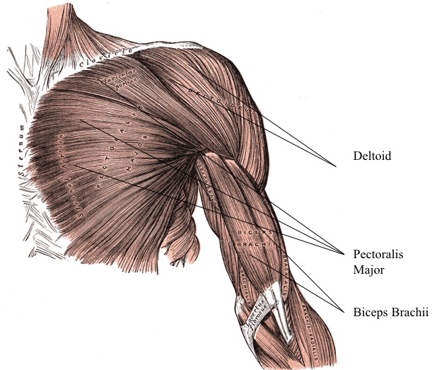
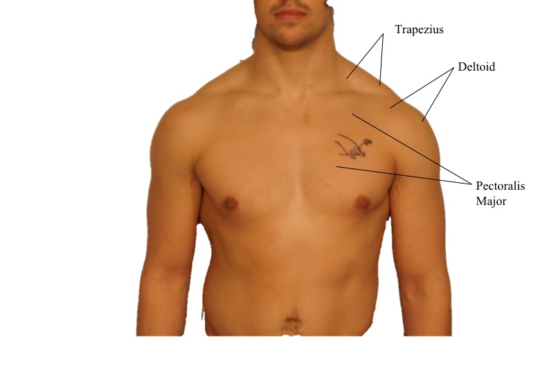
The superficial muscles of the anterior pectoral girdle that are easily identifiable are the Deltoid and pectoralis major:
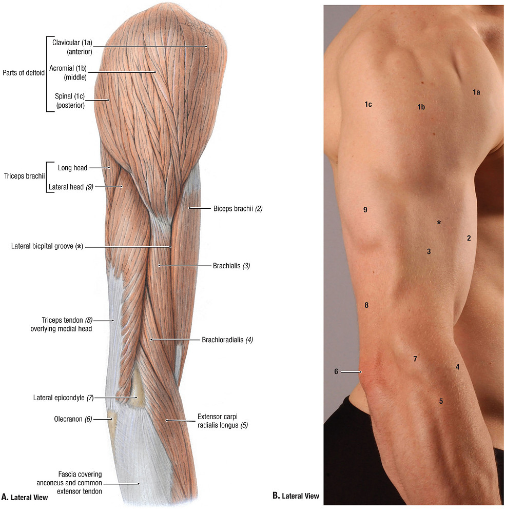
Muscles of The Upper Arm
https://www.youtube.com/watch?v=Lb0B8Ei3pHs
https://www.youtube.com/watch?v=0kT5cUwSnAo
Identifying muscle’s on yourself
Biceps Brachii: These muscles are best seen when the arm is in a supine position with the elbow in flexion. You can feel the biceps brachii tendon at the distal end of the humerus, inserting into the cubital fossa on the proximal end of the forearm.
Triceps Brachii: These muscles can be felt when the arm is extended, more so against resistance.
Surface Anatomy of the Forearm and Wrist
The antebrachium, or rather the forearm, is the region between the elbow and wrist; and, the carpus, or wrist, is between the forearm and palm. By now you’re probably reminded of Stealers Wheel hit song “Stuck in the Middle with You,” and I apologize for it being stuck in your head, or a sudden urge to watch Quentin Tarantino’s Reservoir Dogs (1992). Moving forward, the following are some highlights of the prominent surface anatomy of the forearm and wrist.
Bony Features
- Ulna. Found within the medial aspect of the forearm, this bone can be completely palpated from the olecranon process all the way to the styloid process – a projection at the most distal end of the ulna, toward the little finger. Proximal to the styloid process is head of the ulna, a rather obvious enlargement on the dorsal surface.
- Radius. Unlike the ulna, the radius can be partially palpated with ease, the distal end is an example of such but the proximal end is enveloped in muscles. The styloid process of the radius is a prominent projection on the most distal, lateral edge of the forearm.
- Pisiform bone. Located within the proximal row, and the medial edge of the carpal bones, it can be palpated as a projection distal and anterior to the styloid process of the ulna.
Muscular Features
Isolating the muscles of the forearm is no simple task, but it is simpler to examine the tendons as they approach the wrist and then to move proximally to unveil the musculature:
- Brachioradialis. Located at the most superior and lateral aspect of the forearm.
- Flexor carpi radialis. On the lateral edge of the forearm, and about 1cm medially towards the styloid process of the radius.
- Palmaris longus. Fun fact, only about 15-20% of the population does not have this muscle in at least one arm. For those who do possess this muscle, simply flex the wrist slightly and hold the thumb and pinky together so it can be seen quite prominently.
- Flexor digitorum superficialis. Located medially to the palmaris longus tendon; and, this tendon can also be palpated by flexing the fingers at the proximal interphalangeal and metacarpophalangeal joints.
- Flexor carpi ulnaris. Located on the medial edge of palmaris longus tendon.
Vasculature: Radial Artery
The radial artery is located on the lateral aspect of the wrist between the styloid process of the radius and flexor carpi radialis tendon. This area is commonly used to take one’s pulse; this can be either by supporting the wrist with your palm on the dorsal surface and wrapping your fingers around just beneath the crook of the thumb or just by pressing this surface with the index and middle fingers.
“The Anatomical Snuffbox”
This is an area that is aptly named by its historical significance of individuals with the habit of taking a pinch of snuff (scented powder or powdered tobacco), placing this substance into this area, and then subsequently sniffing it into the nose. The area is a triangular depression between the tendons extensor pollicis longus and brevis. Other features to be palpated in this area include the styloid process of the radius, scaphoid, trapezium, first metacarpal, and radial artery.
Wrist Creases
If you regard the anterior surface of the wrist, there are a series of creases or more-or-less constant lines. Each are aptly named as proximal, middle and distal, and are also attached firmly to the underlying fascia.
Surface Features of the Hand
Dorsal Surface
- Knuckles. Commonly refers to the distal aspects of heads of metacarpals II-V, but also includes dorsal aspects of metacarpophalangeal and interphalangeal joints.
- Dorsal venous arch. This can be seen by compressing the blood vessels at the wrist as the hand opens and closes; they are superficial veins on the dorsum of the hand that drain blood into the cephalic vein.
- Tendon of extensor pollicis brevis muscle. The tendon is in line with the phalanx of the pollex, or rather the thumb.
- Tendons of extensor digitorum muscle. The tendons of this muscle are located on the dorsum of the hand in line with the phalanges of the index, middle, and little fingers.
- Tendon of extensor digiti minimi muscle. This tendon can be seen on the dorsum in line with the little finger.
Palmar Surface
- Thenar eminence. Largest rounded contour on the lateral aspect of the palm, formed by the muscles that move the thumb. This area is also called the “ball of the thumb.”
- Hypothenar eminence. A smaller rounded contour on the medial aspect of the palm, formed by the muscles that move the little finger. Also called the “ball of the little finger.”
- Palmar flexion creases. Skin creases on the palm.
- Digital flexion creases. Skin creases in the anterior surface of the fingers.
