Thorax
Joints, Ligaments and Connective Tissues
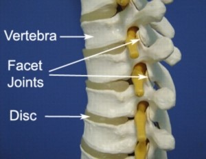
Facet Joint:
Is made of small, bony knobs that line up along the back of the spine. Where these knobs meet, they form a joint that connects the two vertebrae. The alignment of the facet joints of the thoracic spine allows freedom of movement as you twist back and forth or lean side to side. Thoracic facet joints are not as active as the cervical or lumbar facet joints, the thoracic area of the spine is not as prone to damage as other areas of the spine. As the vertebrae continually pivot and move, the cartilage lining on the facet joint can wear down, making the facet joint prone to inflammation and swelling, which could lead to joint stiffness.
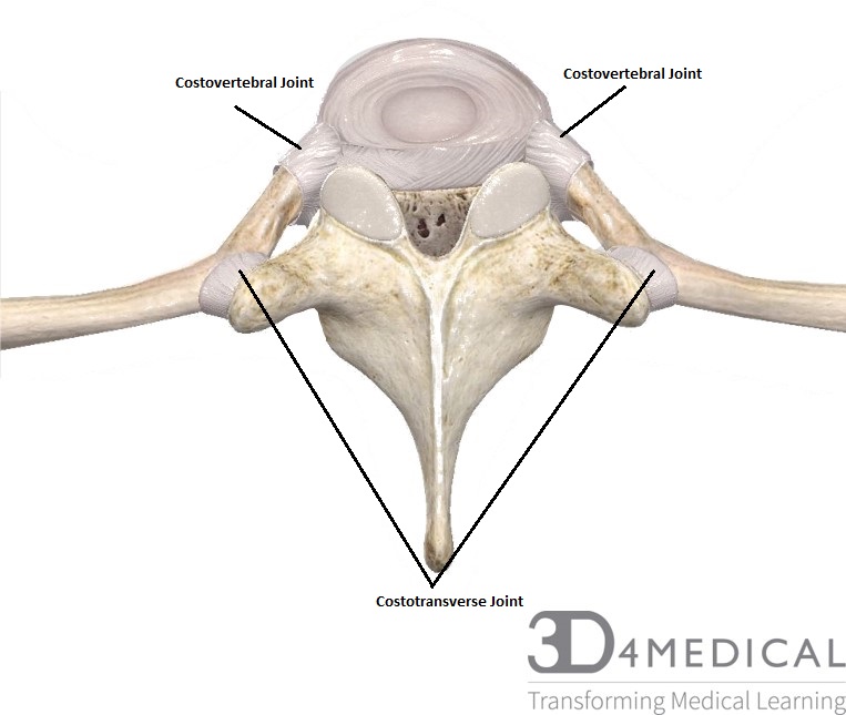
Costotransverse joint:
Is an articulation between the articular costal tubercle of the rib and the costal facet of the transverse process of a thoracic vertebra. The costovertebral joint is the articulation between the costal facets or demi-facets (formed by the caudal side of the superior vertebra and the cranial side of the inferior vertebra) and the head of the rib. These facets form a solid angle whose base consists of the annulus fibrosis of the intervertebral disc. A synovial joint ensures the connection between the rib and the thoracic vertebra. Together with the thoracic cage, the costovertebral and costotransverse joints provide stability.
Ligaments
Anterior Longitudinal ligament:
The anterior longitudinal ligament runs down the ventral surface of the spine from the occiput to the sacrum. The anterior longitudinal ligament is thicker and narrower in the thoracic region. The ligament is thick and slightly narrower over the vertebral bodies and firmly attached to the edges of the vertebral bodies. The ligament has three layers: superficial, intermediate and deep. The superficial layer traverses three or four vertebrae, the intermediate layer covers two or three and the deep layer is only between individual vertebrae. The deep layers of the ligament, as it crosses the vertebral body, blend with the periosteum. The anterior longitudinal ligament provides stability, allow limited mobility and hold the anatomical components of the spine together.
Posterior Longitudinal Ligament:
The posterior longitudinal ligament is situated within the vertebral canal. The posterior longitudinal ligament is composed of smooth, shining, longitudinal fibers, denser and more compact than those of the anterior longitudinal ligament, and consists of superficial layers occupying the interval between three or four vertebrae, and deeper layers which extend between adjacent vertebrae. It limits flexion of the vertebral column and reinforces the intervertebral disc.
Ligamentum Flavum:
Ligamentum flavum is a yellow-colored ligament that connects the vertebrae in the neck and back. This ligament provides protection to the neural elements of the spine and provides stability by preventing excess motion between vertebrae. Ligamentum flavum is the strongest ligament in the spine but its size and shape adapts to the body’s movements. For instance, this ligament stretches to provide the spinal canal with more room when sitting or leaning forward. When standing or leaning back, it provides less room for the spinal nerves because it is shorter and thicker.
Interspinous Ligaments:
Are thin and membranous ligaments that connect adjoining spinous processes of the vertebra in the spine. They extend from the root to the apex of each spinous process. They meet the ligamenta flava in front and blend with the supraspinous ligament posteriorly. The ligaments are narrow and elongated in the thoracic region. The role of the interspinous ligament is to limit flexion (bending forwards) through restricting separation of the spinous processes of the vertebral column.
Supraspinous Ligament:
The supraspinous ligament (aka supraspinal ligament) is a strong, fibrous cord, that connects the tips of spinous processes from the seventh cervical vertebrae to the sacrum. This ligament is continuous with the nuchal ligament superior to C7. This ligament helps to limit flexion of the vertebral column. The supraspinous ligament also helps about as a back stabilizer. This ligament is the most superficial ligament of the five listed above.
