Pelvis
Nerves, Blood Vessels and Lymph
Nerves, Blood Vessels and Lymph of the Gluteal Region
Nerves of the Gluteal Region
Originating from the sacral plexus (L4 – S4), there are a number of significant nerves that either directly innervated the buttocks or pass through the gluteal region to innervate more inferior regions such as the perineum and thigh.
These nerves can be categorized into the superficial (clunial nerves) that provide sensation to the skin of the gluteal region, and the seven deep gluteal nerves (superior gluteal nerve, inferior gluteal nerve, sciatic nerve, nerve to quadratus femoris, posterior cutaneous nerve of the thigh, nerve to obturator internus, and the pudendal nerve) that either directly innervate gluteal structures or continue their course inferiorly to other regions.
Superficial Gluteal Nerves:
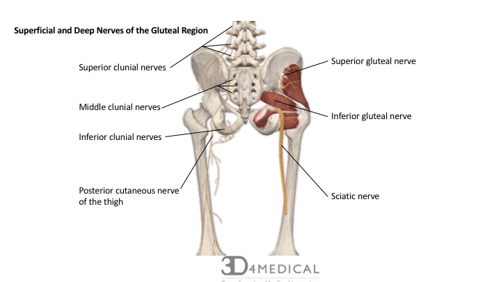
The Clunial Nerves
The most superficial nerves of the gluteal region are the inferior, middle and superior clunial nerves. They provide sensation within the borders of the iliac crest, between the posterior superior iliac spine and the iliac tubercles.
The superior clunial nerve originates from the posterior rami L1 – L3 spinal nerves of the lumbosacral plexus, spans inferolaterally across the iliac crest and supplies the skin of the superior buttocks.
The middle clunial nerves originates from the posterior rami of S1 -S3 spinal nerves of the sacral plexus, travels through the sacral formina and spreads laterally surface to supply the skin of the sacrum and skin close to the intergluteal cleft of the buttocks.
The inferior clunial nerve originates from the posterior rami of S2 – S3 spinal nerves of the sacral plexus, exiting at the inferior border of the gluteus maximus muscle before travelling in a superior direction to supply the inferior half of the buttocks to the level of the greater trochanter.
Deep Gluteal Nerves:
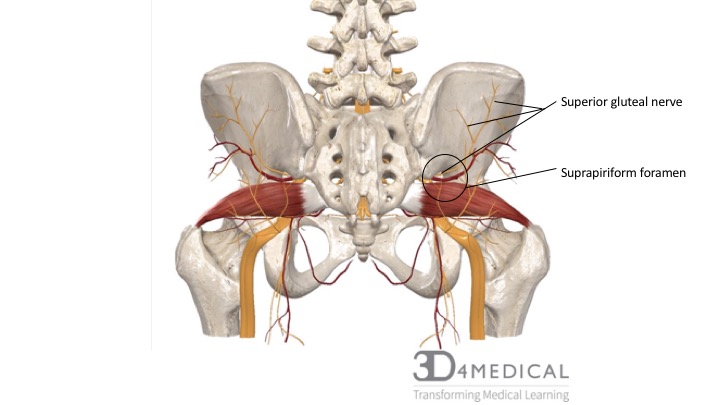
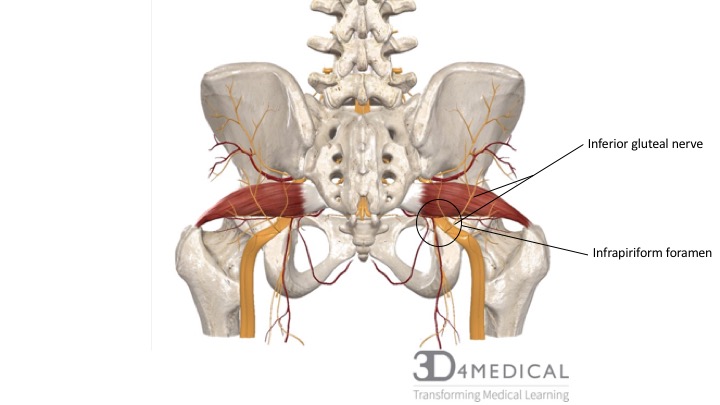
The seven deep gluteal nerves all originate via the lumbosacral plexus or sacral plexus, all exit the pelvic region through the greater sciatic foramen and with the exception of the superior gluteal nerve, all travel through the infrapiriform foramen (inferior to the piriformis) to innervate gluteal regions or other inferior structures.
Superior Gluteal Nerve
The superior gluteal nerve originates from the posterior divisions of anterior rami L4 – S1 spinal nerves of the sacral plexus. It enters the gluteal region via the greater sciatic foramen superior to the piriformis (suprapiriform foramen) and travels in a lateral fashion along-side the deep branch of the superior gluteal artery between the gluteus minimus and gluteus medius muscle. The superior gluteal nerve further branches into a superior branch that supplies the gluteus medius muscle, and an inferior branch that supplies both the gluteus medius, gluteus minimus and the tensor fasciae latae.
Inferior Gluteal Nerve
The inferior gluteal nerve originates from the posterior division of anterior rami L5 – S2 spinal nerves of the sacral plexus. It enters the gluteal region via the greater sciatic foramen inferior to the piriformis (infrapiriform foramen). The inferior gluteal nerve travels closely with the branches of the inferior gluteal artery and inferior gluteal vein and superficially to the sciatic nerve before dividing into multiple branches that course to innervate the superficial gluteus maximus.
Sciatic Nerve
Although the sciatic nerve does not supply any of the gluteal musculature, because of its relationship to the piriformis and individual variability in its course, it is an important structure implicated in the clinical condition known as piriformis syndrome (see Pelvic Clinical Conditions).
Nerve to Quadratus Femoris
The nerve to quadratus femoris originates from the anterior division of anterior rami L4 – S1 spinal nerves of the sacral plexus. Travelling through the infrapiriform foramen to exit the pelvis, the nerve to quadratus femoris courses deep to the sciatic nerve, before passing over the posterior aspect of the hip joint to innervate both the quadratus femoris muscle and inferior gemellus.
Posterior Cutaneous Nerve of the Thigh
The posterior cutaneous nerve of the thigh originates from the anterior and posterior divisions of anterior rami S1 – S3 spinal nerves of the sacral plexus. The posterior divisions of the anterior rami S1 and S2 innervate the skin of the inferior half of the buttocks via the inferior clunial nerves.
Nerve to Obturator Internus
The nerve to obturator internus originates from the posterior divisions of anterior rami L5 – S2 spinal nerves of the sacral plexus. Recalling the unique course of the obturator internus muscle, the nerve travels through the infrapiriform foramen to wrap around the ischial spine where it innervates the superior gemellus, and then courses through the lesser sciatic foramen into the perineum to innervate obturator internus.
The Pudendal Nerves
The pudendal nerve does not innervate any muscles in the gluteal region, but is sometimes included with the deep gluteal nerves because of its path through through the pelvis coursing through the infrapiriform foramen. See below sections for further information on the pudendal nerves.
| NERVES OF THE GLUTEAL REGION | |||
| Nerve | Origin | Course | Innervation |
| Clunial – superior | Posterior rami of L1 – L3 spinal nerves | Travel in an inferolateral direction across the iliac crest | Supply skin of superior buttocks |
| Clunial – middle | Posterior rami of S1 – S3 spinal nerves | Exit via sacral foramina and pass travel laterally to gluteal region | Supply skin over sacrum and adjacent area of buttocks |
| Clunial – inferior | Anterior rami of S2 – S3 spinal nerves | Travels from inferior gluteus maximus border and ascends superficially to gluteus maximus | Supply skin of inferior half of buttocks as far as greater trochanter |
| Sciatic | Sacral plexus L4 – S3 spinal nerves | Enters via greater sciatic foramen inferior to piriformis and deep to gluteus maximus | Supplies no gluteal muscles, but is an important nerve of the gluteal region due to its relationship to piriformis |
| Posterior cutaneous nerve of thigh | Sacral plexus S1 – S3 spinal nerves | Enters via greater sciatic foramen inferior to piriformis and deep to gluteus maximus | Supplies inferior lower half of buttocks via inferior clunial nerves |
| Superior gluteal | Sacral plexus L4 – S1 spinal nerves | Enters via greater sciatic foramen superior to piriformis; travels laterally between gluteus medius and minimus and terminates its course at tensor fasciae lata | Innervates gluteus medius, gluteus minimus and tensor fasciae latae |
| Inferior gluteal | Sacral plexus L5 – S2 spinal nerves | Enters via greater sciatic foramen inferior to piriformis and deep to inferior gluteus maximus | Gluteus maximus |
| Nerve to quadratus femoris | Sacral plexus L4 – S1 spinal nerves | Enters via greater sciatic foramen inferior to piriformis, deep to sciatic nerve | Hip joint, inferior gemellus, and quadratus femoris |
| Nerve to obturator internus | Sacral plexus L5 – S2 spinal nerves | Exits pelvis via greater sciatic foramen inferior to piriformis; descends posterior to sacrospinous ligament | Supplies superior gemellus and obturator internus |
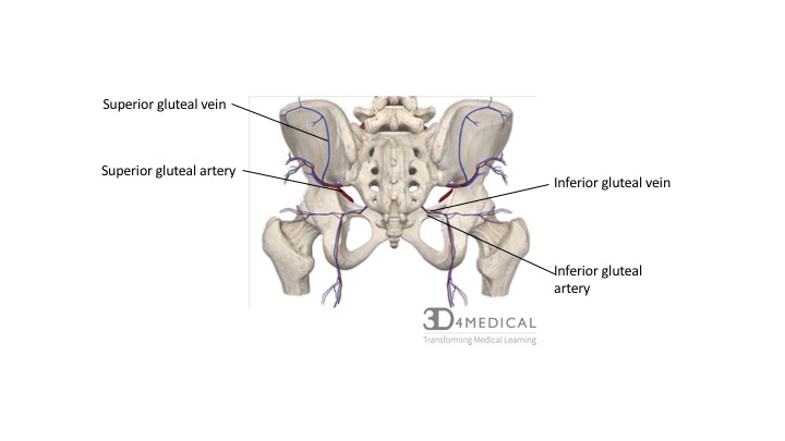
Blood Vessels of the Gluteal Region
Arteries of the Gluteal Region
The major arteries that supply the gluteal region are the superior gluteal artery and the inferior gluteal artery. Both of these arteries bifurcate via the internal iliac arteries.
The largest branch to bifurcate from the internal iliac artery, the superior gluteal artery divides further after its course through the suprapiriform foramen into a superficial branch that supplies the gluteus maximus and an inferior branch that supplies the gluteus minimus, gluteus medius and the tensor fasciae latae.
The inferior gluteal artery also originating via the internal iliac arteries, descends through the infrapiriform foramen travelling deep the gluteus maximus and medial to the sciatic nerve. This artery is responsible for supplying the gluteus maximus, obturator internus, quadratus femoris, and proximal fibers of the hamstrings.
Veins of the Gluteal Region
The superior and inferior gluteal veins arise from the internal iliac veins and parallel the course of the superior gluteal artery and inferior gluteal artery through the suprapiriform foramen and the infrapiriform foramen to drain blood from the gluteal region. As a safety mechanism the gluteal veins connect with femoral vein tributaries to provide an alternate path for blood return if the femoral vein becomes occluded.
Nerve Innervation of the Pelvic Floor
The pelvic floor muscles are innervated through somatic, visceral, and central pathways. The pudendal nerves pass through the pudendal canal. The main nerve that innervates the pelvic floor is the pudendal nerve that arises from the ventral branches S2-S4 of the sacral plexus. The pudendal canal is located through the lateral wall of the ischiorectal fossa. The nerves and vessels are surrounded by a fascia sheath. The pudendal nerve is derived from the second, third, and fourth sacral spinal nerves. The nerve exits the pelvis through to the gluteal region between piriformis and coccygeus through the greater sciatic foramen and crossing through the sacrospinous ligament close to the attachment spot to the ischial spine. It then divides into the dorsal and perineal nerve of the penis or clitoris.

Blood Supply of the Pelvic Floor:
Arteries of the Pelvic Floor
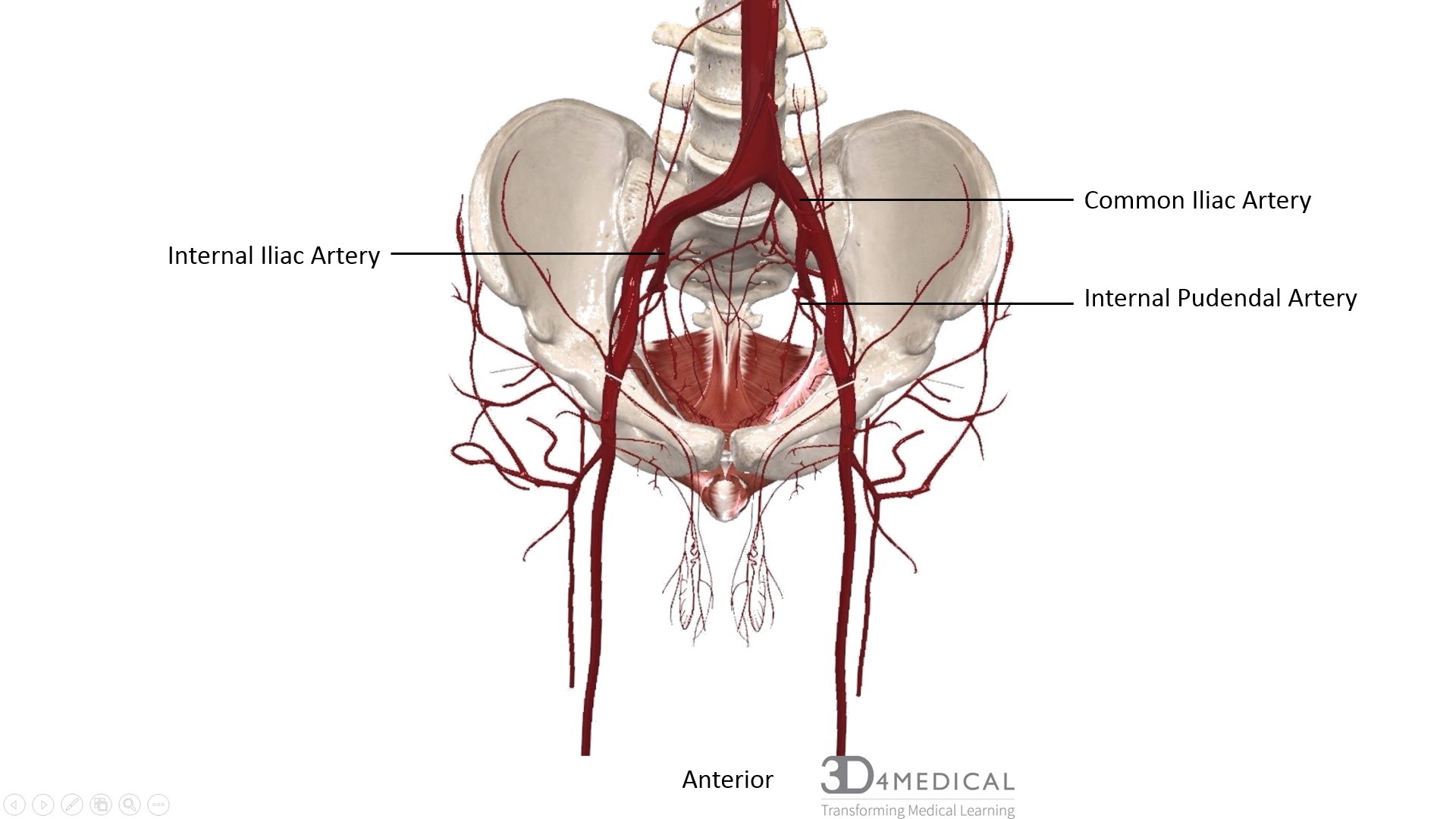
The pelvic floor is supplied by the common iliac artery that branches from the aorta. The common iliac artery supplies the pelvic organs and the lower limbs. The common iliac artery then branches into the external iliac artery and internal iliac artery which supplies the pelvic organs, pelvic walls, buttocks, medial thighs and perineum. The internal pudendal artery is a branch from the internal iliac artery that leaves the pelvic cavity and supplies the perineum and pelvic floor.
Veins of the Pelvic Floor
The pelvic floor is drained by the veins of the pelvic floor, including the external iliac vein, the internal iliac vein and the common iliac vein. These three veins correspond with the three major pelvic artier. A condition referred to as pelvic congestion syndrome occurs when blood builds within the veins of the pelvic region (refer to Pelvic Clinical Conditions).