Thorax
The Dorsal Cavity
The Dorsal Cavity
This section of the textbook will highlight the central nervous system, focusing on the structure and function of the lower brain stem and spinal cord. It will summarize the unique features of the vertebrae, the major supporting muscles and the ligaments that provide stability and strength for the spinal column.
Introduction
The dorsal cavity is the entirety of the back; it consists of the brain stem and the entire spinal cord. It also consists of all the nerve plexuses and single nerves that exit the spinal cord via the spinal column. Additionally, the dorsal cavity is comprised of all the muscles and ligaments that make the back mobile and strong. The function of the dorsal cavity gives rise to sensation and perception of the nervous system. The nerves originate at the spinal cord, then travel long distances to innervate different regions of the body. The central nervous system, being that of the brain and spinal cord, controls all body functions; from organ control to movement. Most importantly, the central nervous system acts as a vessel to communicate stimulation in the brain to actions and reactions within the body. To continue to explore the phenomena of the central nervous system and dorsal cavity, we must first discuss the structure that encompasses and protects the brain stem and spinal cord: the vertebrae.
The Vertebrae—spinal curves
The bones that make up the vertebral column are complex but serve a great purpose to protect the spinal cord and the corresponding roots that make up the central nerve system. The vertebrae consist of four different regions; the cervical vertebrae, which are smaller in size and support the head; the thoracic vertebrae, which are larger in size and provide rib attachments; the lumbar vertebrae, which are very large and support most of the upper body. Finally, the sacrum vertebrae are the last bones of the spinal column that are completely fused together. Let’s delve a little deeper into the characteristics of these unique bones.
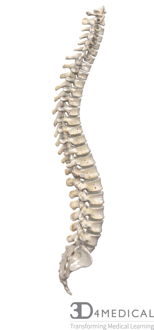
Spinal Curves
The Cervical Vertebrae:
These vertebrae are petite in size and provide support for the entire head, there are seven cervical vertebrae in total. They have a very large vertebral foramen and they have transverse foramina in the transverse processes to supply space for veins and arteries to the brain. The cranium balances, quite precariously on the Atlas (C1), pivoting with the occipital condyles of the inferior portion of the occipital bone. Based on the structure of the Atlas, it allows the head to nod in the “yes” motion. The Axis (C2), allows the head to rotate and provides the “no” motion of the head. There is no intervertebral disc between Atlas and Axis, where there are usually in the rest of the vertebrae. The vertebra prominens (C7), is also a special landmark of the spine; it has a long spinous process with a tubercle that makes it rather bulky, therefore, it protrudes in the back, at skin of the neck. C7 is the transition point between the cervical vertebrae and the thoracic vertebrae. These vertebrae are capable of lots of movements, such as rotation, lateral bending, flexion and extension.
The Thoracic vertebrae:
The thoracic vertebrae are larger in size and have a heart shaped body. They have a round shaped vertebral foramen, and long slender spinous processes that slant inferiorly. A unique feature of these vertebrae is that they have coastal facets for rib attachment. The thoracic region of the spine is a little more limited in movement; rotation occurs here, lateral bending and flexion and extension; however, not to the same extent as the cervical or lumbar regions. Kyphosis is a spinal disorder that occurs when a posterior exaggeration occurs after continuous flexion or forward motion in this region.
The Lumbar vertebrae:
These vertebrae are the largest in the vertebral column; they provide support for the entire upper body. They are oval shaped and have a triangular vertebral foramen; they have short spinous and transverse processes and their articular facets are vertical and face medially and laterally. More movement occurs here such as flexion and extension, rotation and lateral bending. This region of the spine is prone to lordosis, a disorder in the spine where there is an anterior exaggeration in the vertebrae. It occurs naturally sometimes for pregnant women with the increasing weight of the infant in the belly; however, if lordosis proceeds or continues to get more defined, then nerve impingement and other nerve system dysfunctions can arise.
The Sacrum
The sacrum is the caudal end of the vertebral column, it provides places for muscle attachment among other things. They are the 3-5 fused vertebrae in the vertebral column that attach to the pelvic girdle. They fuse during puberty and between the ages 25-30yrs old. They have no movement, but merely provide structural support and protection of the pelvic floor.
Joints
The intervertebral discs provide most of the support during articulation between the vertebrae. The inferior articular processes articulate with the superior articular processes of the more caudal vertebrae. The joints in the vertebrae allow the spine to move in a variety of ways through extension, flexion, lateral bending and rotation. Over time, the intervertebral discs can become impinged. Many individuals complain about back pain, especially lower back pain; this can occur from deteriorating discs, or when the discs compress. See below for more information on the ligaments and connective tissue in this region.
Muscles
| Errector Spinae | Spinalis | Longissimus | Iliocostalis | Quadratus lumborum | Multifidus |
| capitis | Longissimus capitis | ||||
| cervical | Spinalis cervicis | Longissimus cervicis | Iliocostalis cervicis | ||
| thoracic | Spinalis thoracis | Longissimus thoracis | Iliocostalis thoracis | ||
| lumbar | Iliocostalis lumborum | ||||
For more information see the thoracic region section of text.
Intervertebral Muscles
| Rotators | Longus capitis | Intertransversarii |
| Interspinales | Longus coli |
See below for more information on the origins and insertions on these muscles in the lumbar region (they span the whole spine).
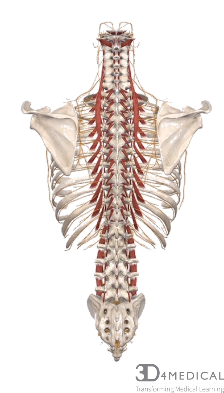
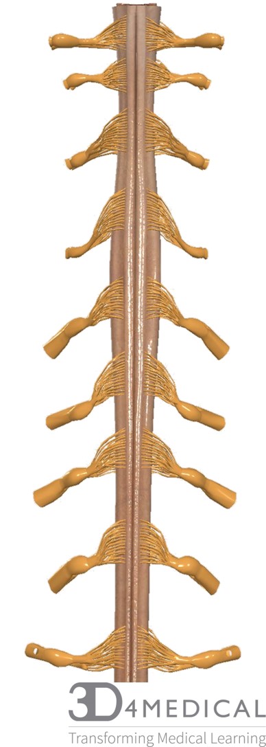
Full spine with intrinsic muscle attachments and nerves
Blood
The vertebral arteries supply the brain and spinal cord with blood from the heart. They branch off the subclavian which supplies first and foremost the shoulders, arms, back and central nervous system. For more information on the blood supply, refer to the cervical vertebrae section where there is more detail.
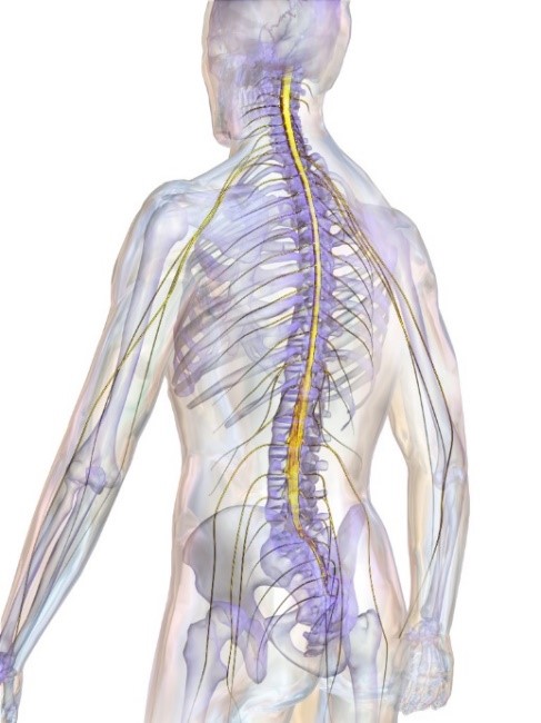
Nerve Supply
The central nervous system (CNS) is a very important structure to consider when discussing the dorsal cavity. The nervous system is the first system to develop in the fetus. It plays a specific role in the ability for us, humans, to actively sense our surroundings, how else are you to perceive your world without the nerve system?
(Wikimedia commons, 2018)
Nerve tissue
The nervous system is a complex structure, in which we must first discuss what makes up neural tissue. There are two types of cells that are present in neural tissue; one being neurons that send and receive electrical impulses. The other being neuroglia or glial cells, which are the “helping” cells to the neurons, in which they protect and support the functions that the neurons undertake. There are plenty of neurons; however, there are many more glial cells to neurons. The reason for the substantial amount of glia in the nerve tissue is to aid in the reconstruction of itself after trauma. Neuroplasticity is the ability of the nervous system to rebuild or regrow if part of the brain had to be removed or if certain neurons lost function due to a concussion or stroke. Schwann cells are a type of glial cell that repairs damaged neurons by wrapping around the axons in the peripheral nervous system. It is something that more and more researchers have been investigating. The ability for the brain to improve its own function is astounding! Think about how much information it can retain, especially when learning a new subject in school; the building of new neural pathways is what the nervous system thrives in.
The Central and Peripheral Nervous Systems.
Furthermore, the nervous system can be divided into two subsections: the central nerve system (which this article will focus on) and the peripheral nerve system. The central nerve system consists of the brain and spinal cord. The peripheral nerve system consists of all the receptors as well as all the peripheral nerves in the body.
The central nerve system not only consists of the brain and spinal cord, it also consists of neural and connective tissue as well as all the blood vessels that supply the nerve system. The function of the CNS is to process as well as coordinate sensory data entering and exiting the body, motor commands and organ activity. The CNS is also extraordinarily adept in processing and coordinating higher brain functions such as intelligence, learning, memory and emotion. Within neural tissue, there are action potentials running every second to help us input and output information. Action potentials are created due to the difference in concentration gradient between sodium and potassium ions in the neurons; simply put, it is a difference in membrane potential. These action potentials can import an electrical impulse through the body. There are three types of neuron structures: anaxonic, which have numerous dendrites but no axon; bipolar neurons, which has one dendrite and one axon; unipolar neurons, in which the dendrite and axon are continuous with each other; and finally, multipolar neurons, which have two or more dendrites and a single axon. The CNS has the ability to innervate all different organs and muscles in the body. For example, the nerves in the cervical plexus can innervate muscles and organs in the lower abdominal cavity, such as the phrenic nerve (C3–5) controls the function in the diaphragm supporting lung activity.
The Somatic and Autonomic Nervous System
Something to add, the somatic nervous system (SNS) controls voluntary and involuntary skeletal muscle contractions. The autonomic nervous system (ANS) controls subconscious actions and contraction of smooth muscle and cardiac muscle as well as glandular secretions. The ANS then divides into the sympathetic nerve system, which is stimulating in nature, and parasympathetic nerve system, which is relaxing in nature. The sympathetic and parasympathetic are never on and off; rather, they are working all the time at different rates, called “sympathetic tone”. All these qualities of the nervous system help it strive and provide for the body. Without the nerve system, it would be impossible to have a connection with our environment.
Visceral Organs: these organs keep us alive
There are many visceral organs in the body that play acute roles in keeping the body alive. This section will describe the importance of the brain stem and spinal cord in providing the body with sensation and perception. The nervous system is the ultimate sensory processing tool in the body! Without these structures in the brain stem and spinal cord, the body would have no way of actively processing sensory information into higher levels of intelligence. Let’s take a closer look at these structures.
The pons relays sensory information to the higher processing centers: the cerebellum and the thalamus. The pons specifically plays a role in subconscious somatic and visceral motor centers. The pons connects to the medulla oblongata which has the task of relaying sensory information to the thalamus and to various places in the brain stem. Additionally, it provides an autonomic center for regulation of visceral functions such as the respiratory, cardiovascular and digestive activities that occur in the body without extra thought. The spinal cord is naturally a visceral organ that relays sensory information and keeps the body moving and functional; without the spinal cord there would be no connection between the mind and body. In other words, there would be no way for the brain to receive sensory information that the body receives with its external environment.
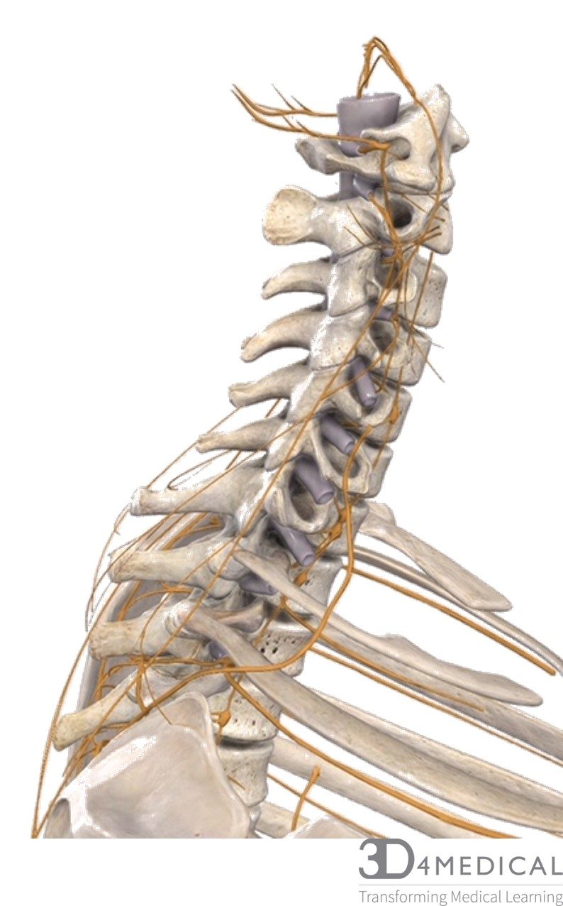
Cervical nerves close up; lateral view
The Spinal Cord
The spinal cord is a significant part of the CNS; it relays sensory information from the body to the brain, and likewise, intellectual information from the brain to the body. It has three main layers that keep it healthy and functioning. The pia mater which is delicate and protects the deep nervous tissue, the arachnoid mater which resembles a spider web, and the dura mater which is the outside portion of the spinal cord. In general, the maters are the meningeal layers that protect the spinal cord and improves its function overall.
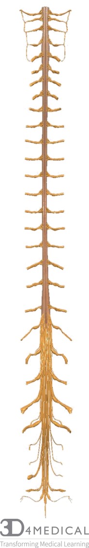
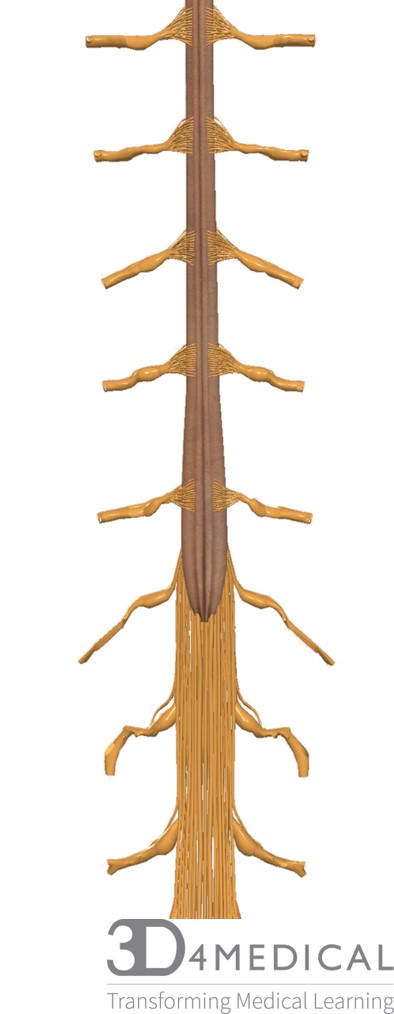
Full spinal cord and its roots (posterior view)
Spinal Nerves
This section will discuss the spinal nerves exiting the spine; it will first focus on the cervical plexus (see table below). Keep in mind that some of these nerves stay local, and others travel long distances in the body. This section will also consider different nerve root pathologies, in which how they occur and how to treat them. Additional to the cervical plexus is the brachial plexus, which branches off C5-C8 and T1. See the brachial region of the text for a closer look at the brachial plexus.
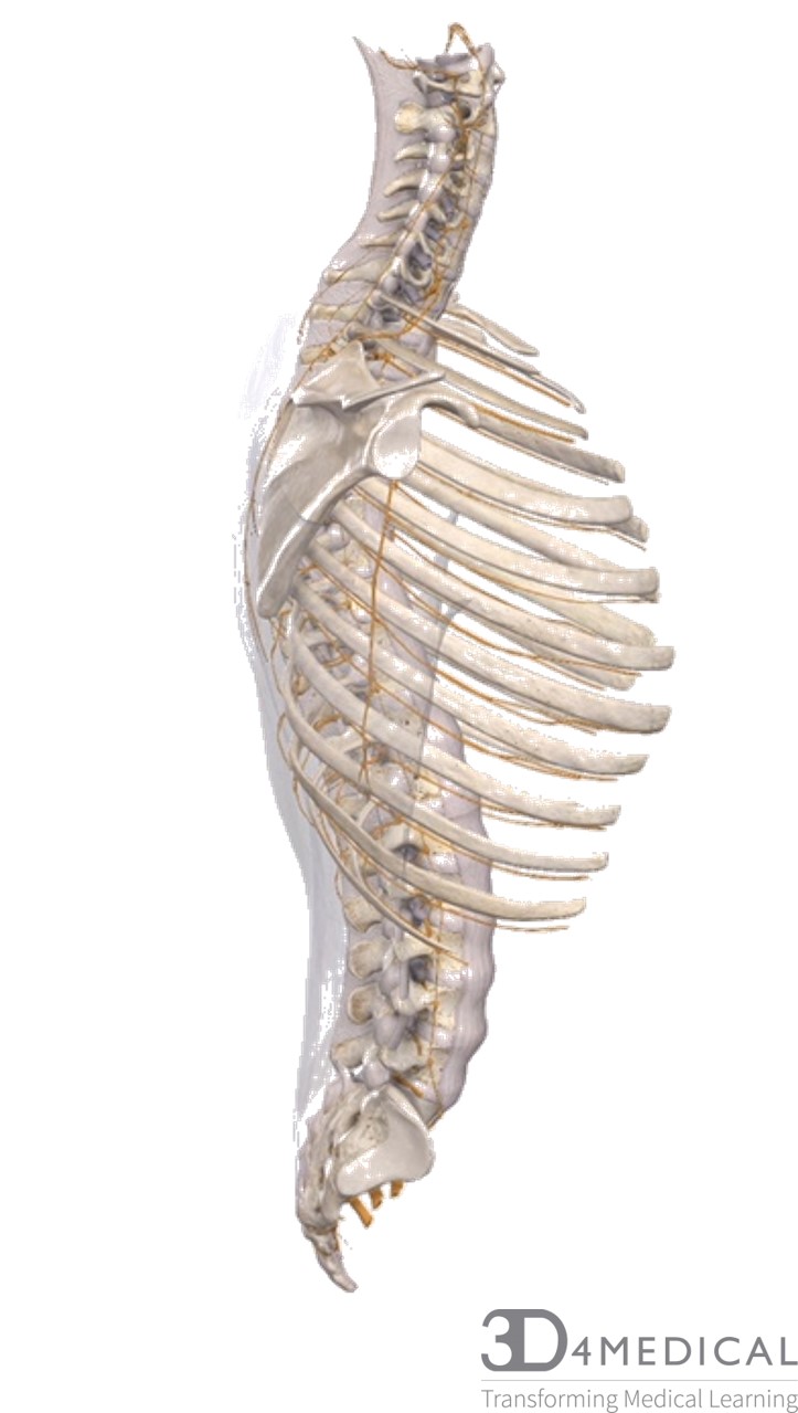
Full spine with spinal nerves and connective tissue (lateral view)
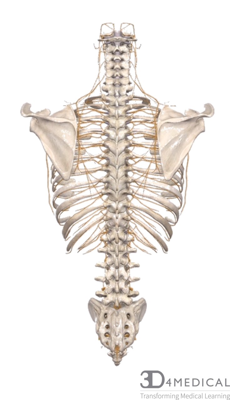
Full spine and spinal nerves (posterior view)
Table of the Cervical Plexus Nerves:
| C1-C4 | C2 | C2-C3 | C3-C4 | C3-C5 |
| Superior/ inferior Ansa Cervicalis
(innervates the five extrinsic laryngeal muscles by Cranial nerve XII) |
Lesser occipital nerve | Great Auricular nerve | Transverse cervical nerve and supraclavicular nerve | Phrenic nerve |
|
Innervates the skin of the neck and scalp superior and posterior to the ear. | Innervates the skin in the posterior aspect of the ear and neck. | The transverse innervates the anterior triangle of the neck. The supraclavicular innervated the skin of the neck and shoulder. | Innervates the diaphragm |
Youtube Drawing of cervical plexus
(fatcat2983472, 2018).
The cervical plexus and the spinal cord in this region have a very critical role in the body. Although the spinal cord in whole has great relevance, it is worth taking a closer look at the cervical area. This is where the connection between the brain stem and the spinal cord occurs. The brain stem is key in providing the body with autonomic function, such as keeping the body standing upright, and processing information. The cervical plexus, in which the spinal nerves exiting the cervical spine, innervate muscles of the neck as well as organs of the trunk including the diaphragm. The last three nerves of the cervical nerves innervate the eyes, salivary glands and heart, C3, C4 and C5 all innervate the diaphragm.
Cervical Enlargement (posterior view)
Thoracic Nerves
The thoracic nerves innervate many muscles; the first six innervate those of the ribs and upper thorax, the lower six thoracic nerves innervate the internal abdominal cavity. To summarize:
| T1 -T3 | T4*, T5 -T8 | T9 -T11 | T12 |
| Heart | Liver | Large Intestine | Large Intestine |
| Lungs | Gallbladder, Stomach, spleen and pancreas | Small Intestine |
*T4 plays a small role innervating these organs
Lumbar enlargement of spinal cord (posterior view)
Lumbar Plexus
The lumbar plexus L1-L4 consists of all the lumbar nerves exiting the lower end of the spinal cord. The L1spinal nerve lies in the L1-2 intervertebral foramen and the L2 spinal nerve lies in the L2-3 intervertebral foramen and so forth. The spinal nerves divide into larger ventral rami and a smaller dorsal ramus. The spinal nerve roots distribute their fibers directly into the ventral and dorsal rami without forming a complete spinal nerve. The lumbar plexus is a bundle of nerves that supplies mostly the lower limbs and pelvic floor. The spinal cord terminates between L1-2 or sometimes as low as L2-3.
Table of Lumbar Plexus Nerves
| T12-L1 | L1 | L1 -L2 | L2 -L3 | L2 -L4 |
| Iliohypogastric nerve | Ilioinguinal nerve | Genitofemoral nerve | Lateral femoral cutaneous nerve | Femoral nerve and Obturator nerve |
| Innervates the external and internal oblique as well as the transverse abdominis muscles. Also innervates the skin over the inferior abdomen and buttocks. | Innervates the abdominal muscles in accordance with the iliohypogastric nerve. Distributes over the skin of the superior thigh and portions of external genitalia. | Distributes over the anteromedial thigh and portions of the external genitalia. | Distributes on to the skin over the posterior, anterior and lateral thigh. | The femoral nerve innervates the quadriceps, sartorius, pectineus and iliopsoas muscles ad well as the skin of the thigh and medial surface of the legs and feet.
The obturator innervates the gracilis, adductor magnus, brevis and longus muscles. It distributes to the skin of the medial surface of the thigh. |
Lumbar Plexus
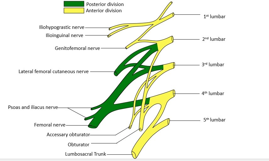
(Wikimedia commons, 2018)
Cauda Equina “Horse’s Tail”
The Latin meaning for cauda equina is horse’s tail; the reason for this is that the spinal cord has already terminated in the lumbar vertebrae, and it is now the remnant dorsal and ventral roots of the spinal segments L2 to S5 that extends inferiorly. In gross dissection, it resembles a horse’s tail. They have a specific function in providing sensation in the pelvic floor and hip girdle. The S1, S2, S3 nerves all play a role in suppling the multifidus muscle in the back and lateral cutaneous branches to the skin and fascia over the sacrum and part of the gluteal region. The lower back, in general, tends to be more susceptible to nerve impingement and back pain; this is because there is more weight and strain the lower back endures. Cauda equina syndrome is a neurological dysfunction that can come about when S1,2,3 are compressed causing lack of sensation in the saddle as well as compromise to the bladder and bowels.
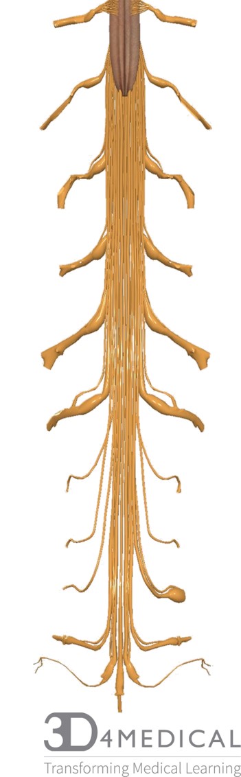
Cauda Equina (posterior view)
Clinical conditions:
Nerve Impingement and Spinal Disorders
As previously mentioned, there are a variety of spinal issues that can occur with the daily stresses of life; such as kyphosis and lordosis, compressed intervertebral discs, and impinged nerves. Scoliosis is a very common distortion in the spine where there is an exaggerated lateral curve in more than one of the movable vertebrae. Most times, it is due to an abrupt growth spurt in an adolescent. Other times it can arise from developmental issues, in which the vertebral bodies are damaged.
Spina bifida is a condition where there is a failure of the vertebral laminae to bond during development. This issue is correlated to developmental abnormalities of the spinal cord and brain; therefore, it is critical to get checked for spinal issues at a young age. One way to test spinal and nervous system health is to perform spinal reflex tests, such as ones when we go visit our doctor and he/she taps on our knee to see if we kick. Reflexes show that there is adequate communication between the body and the spinal nerves. In short, health care practitioners will check different spinal reflexes in the body to determine whether there is an issue in the nervous system or spine.
Health of the Central Nerve System
The CNS is quite a complicated system, so intricate that it is sometimes difficult to understand how it works in harmony with all the activities going on. Some of you may be asking “is there any way to maintain health within the nerve system and spine, since the daily stresses of life have so many effects?” and the answer varies with each professional you encounter. Chiropractic is a profession focussing on the health of the human spine and nerve system. It is a modality of healthcare that does not use drugs or surgery for the treatment of back pain. Having regular appointments with your chiropractor can improve nervous system function and can reduce musculoskeletal disorders. A chiropractor works by making gentle, often manual, adjustments to the spine to correct one or more movable vertebrae that is out of place interfering with the nervous system. A vertebral subluxation is a slight dislocation or dysfunction in the spine and nerve system. Physiotherapists are also admirable professionals that can assist in supporting spinal health, in which they will often prescribe exercises or stretches to the individual suffering from back pain. Therefore, there are a variety of professions that will specialize in supporting the health of the CNS.
In conclusion, the importance of maintaining nervous system health is quite immense in that exercise, yoga, diet and regular appointments with a chiropractor, an osteopath or physiotherapist can truly help or extend the amazing function of this body system. It is a growing research, where there continues to be evidence that the central nervous system is the cornerstone of all body function and development.
References
Chiropractic Association of Australia. https://chiropractors.asn.au/about- chiropractic/chiropractic-and-you (last accessed April 16, 2018).
fatcat2983472. [Youtube] Cervical Plexus Drawing SO GOOD!!!. Available from: http://www.youtube.com/watch?v=Oj9J9b8FIIg (last accessed April 16, 2018).
Martini, F. H., Nath, J.L., & Bartholomew, E.F. (2014). Fundamentals of Anatomy and Physiology (10thed.). Toronto: Pearson.
Physiopedia website. https://www.physio-pedia.com/home/ (last accessed April 16, 2018).
Wikimedia commons website. https://commons.wikimedia.org/wiki/Category:Spinal_cord (last accessed April 16, 2018).
3D4Medical (last accessed March 27, 2018) retrieved from https://3d4medical.com/ [used as an App].
