The Head and Neck
Nerves, Blood Vessels and Lymph
Blood to the Brain
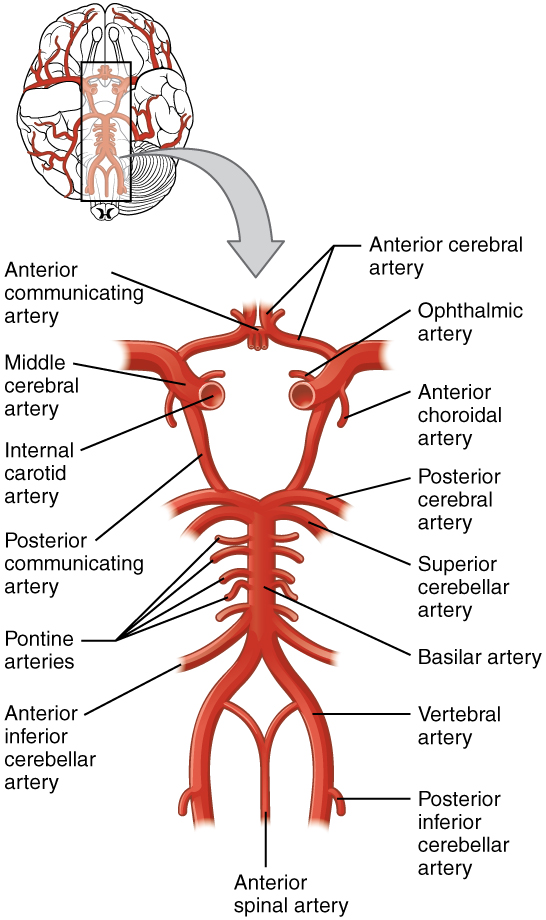
A lack of blood supply to the brain can be detrimental to its functions. Cerebral hypoxia, the lack of oxygen to the brain, can deprive the brain of oxygen and nutrients which can cause it to not function properly. The brain, however, has a system that supplies blood via a network of arteries; therefore, if something were to happen to cut off oxygen and nutrients to a specific artery such as a clot, blood would still flow to other areas of the brain via other arteries.
Blood supply to the brain is supplied by two main pairs of arteries, the internal carotid arteries and the vertebral arteries. The internal carotid arteries are branches of the common carotid arteries and vertebral arteries arise from the subclavian arteries. The common carotid artery goes up the neck and divides into the internal and external carotid arteries. The internal carotid artery then enters the cranium through the carotid canal in the temporal bone. The brains anterior region is supplied by the internal carotid arteries as it reaches most of the cerebrum and the posterior circulation is fed by the vertebral arteries. The vertebral arteries ascend through the cervical vertebrae through the transverse foramina and enter the cranium through the foramen magnum of the occipital bone. The vertebral arteries supply the cerebellum, brain stem, and the underside of the cerebrum.
The two systems connect to form the Circle of Willis. After passing through the skull, the vertebral arteries join to form the basilar artery. The basilar artery and the internal carotid arteries “communicate” with each other via the anterior communicating artery and posterior communicating artery in the Circle of Willis. The communication between the internal carotid and vertebral-basilar systems is an important safety feature of the brain. If one of the major vessels becomes blocked, it is possible for blood flow to come across the Circle of Willis and prevent brain damage.
Venous Return
The venous blood leaves the cranium via the internal jugular veins. They drain the dural sinuses, which are large collections of veins within the dural folds of the dura mater. Within the falx cerebri fold is the superior sagittal sinus and the inferior sagittal sinus. Within the tentorium cerebelli fold, there is the transverse sinus.
Cranial Nerves
| Mnemonic | ||||
| Cranial Nerve Number | Cranial Nerve | Function | Name | Function
M=motor; S=sensory; B=both |
| I | Olfactory | Sense of smell | Oh | Some |
| II | Optic | Sense of sight | Oh | Say |
| III | Oculomotor | Eye movement | Oh | Marry |
| IV | Trochlear | Eye movement | To | Money |
| V | Trigeminal | Face: sensory, motor | Touch | But |
| VI | Abducens | Eye movement | And | My |
| VII | Facial | Face: expression, sensory | Feel | Brother |
| VIII | Vestibulocochlear | Hearing and balance | Very | Says |
| IX | Glossopharyngeal | Tongue and throat: motor, sensory | Good | Big |
| X | Vagus | Parasympathetic | Vegetables | Brains |
| XI | Accessory | Head, neck, shoulders: movement, swallowing | Ah | Matter |
| XII | Hypoglossal | Speech, chewing, swallowing | Heaven | More |
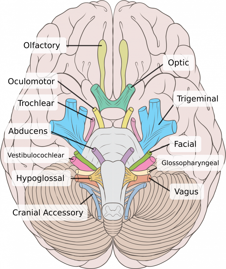
There are twelve pairs of cranial nerves connected to the brain. Cranial nerves are classified based on function. There are four categories: sensory, special sensory, motor, and mixed nerves.
Sensory nerves carry somatic sensory information, such as touch, temperature, pressure, vibrations, and pain. Special sensory nerves carry information like sight, smell, hearing, and balance. Nerves like the olfactory nerve, optic nerve, and vestibulocochlear nerve are special sensory nerves. Motor nerves are axons of somatic motor neurons. This includes nerves like the oculomotor, trochlear, abducens, accessory, and the hypoglossal nerves. Mixed nerves include a mixture of sensory and motor nerves such as the trigeminal, facial, glossopharyngeal, and the vagus nerve.
This is where the face will be discussed…
NERVES
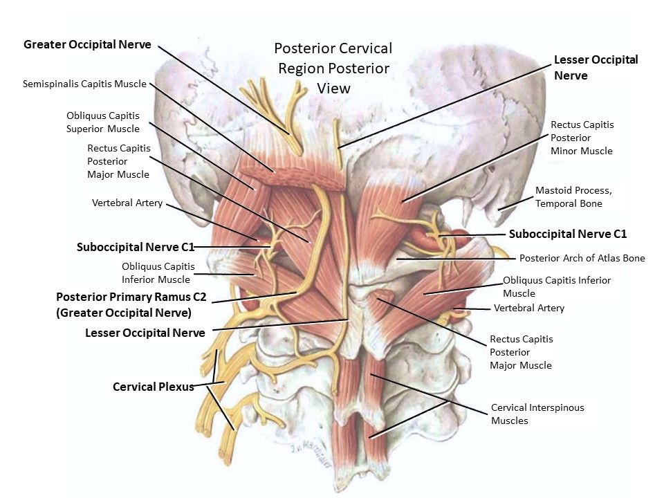
Nerves of the posterior cervical region are primarily branches from the cervical spine nerve rami including the cervical plexus (a branching network of nerves) and some of the roots for the brachial plexus. The nerves in this region innervate muscles in the region as well as the cutaneous parts of the neck and scalp. The eleventh cranial nerve, the Accessory Nerve (CN XI), is also important to this region as a motor nerve to the trapezius muscle and sternocleidomastoid.
Of the Occipital Nerves, the Lesser Occipital Nerve (also known as the Third Occipital Nerve) is closest to the midline and innervates the trapezius, semispinalis and up to the lower part of the scalp.
Lateral to the Lesser Occipital Nerve is the Greater Occipital Nerve that also innervates the semispinalis along with the multifidus and meningeal branches to the posterior cranial fossa. Most lateral is the Lesser Occipital Nerve that provides cutaneous innervation to the posterior surface of the auricle (external ear) and adjacent scalp. The dorsal division is the suboccipital nerve.
NERVES OF THE ANTERIOR CERVICAL REGION
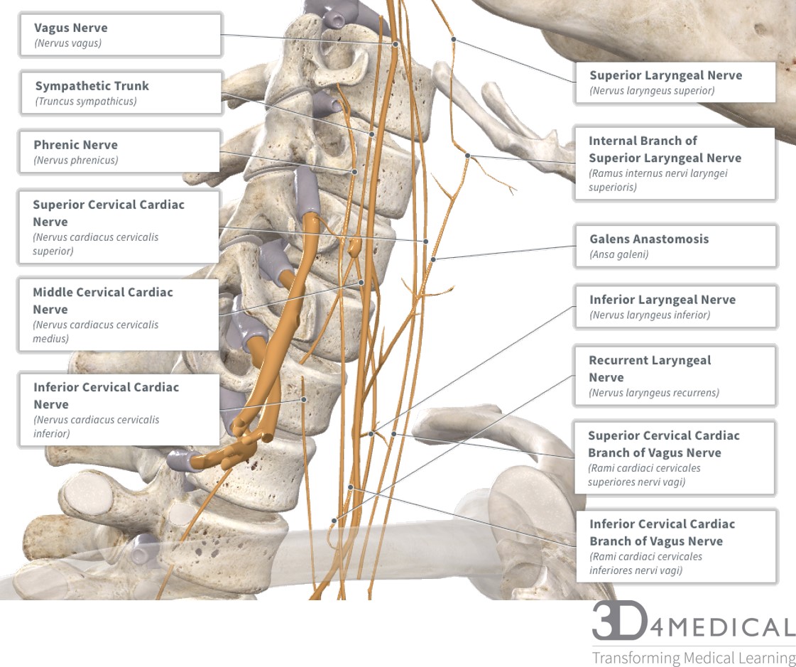
This region has many nerves passing through it. Anterior to the cervical plexus there are 7 nerve branches that pass through this region: Vagus, phrenic, superior cervical cardiac branch of vagus nerve, inferior cervical cardiac branch of vagus nerve, superior cervical cardiac nerve, middle cervical cardiac nerve, and inferior cervical cardiac nerve.
Feature Nerve
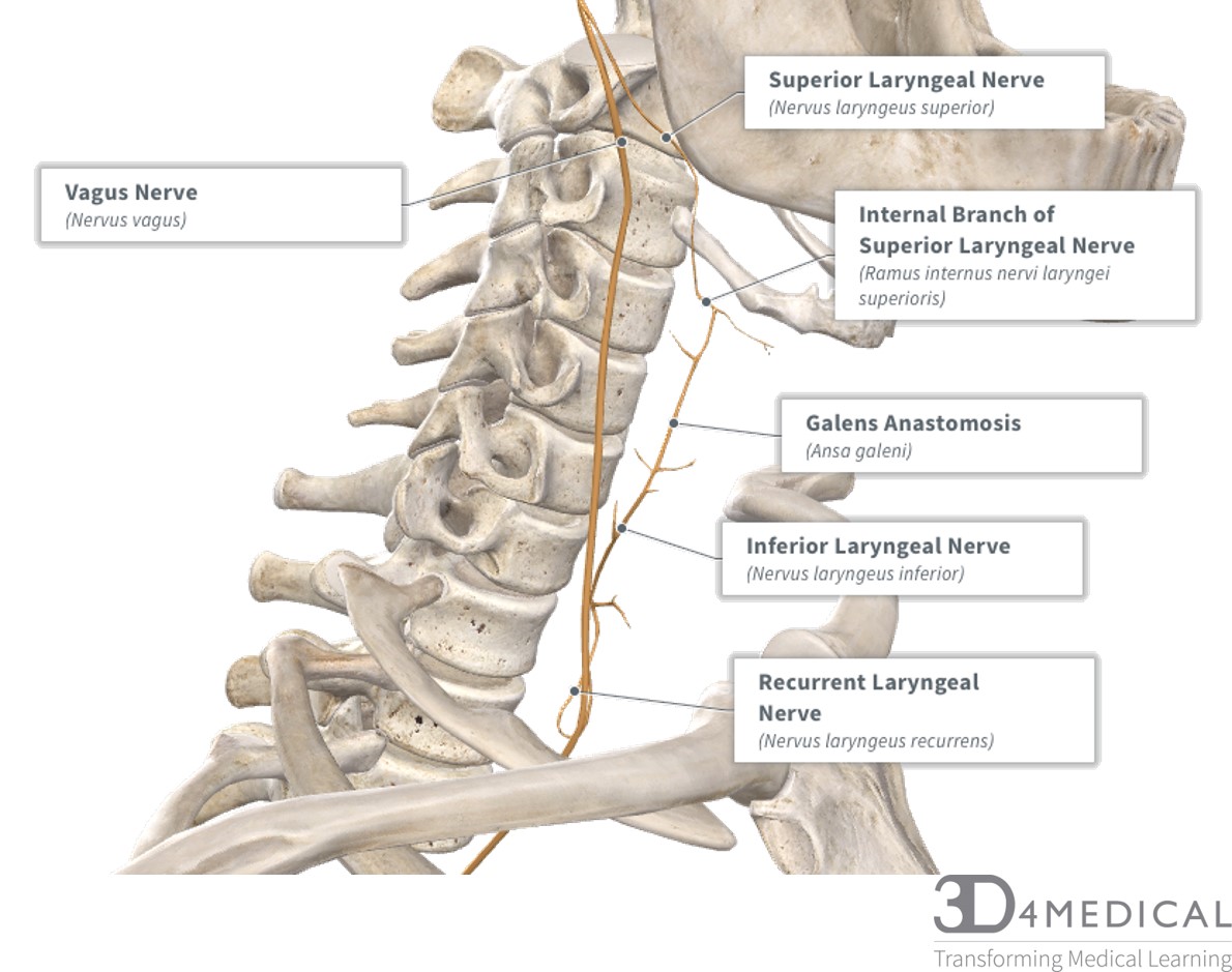
What creates Galen’s Anastomosis?
The superior laryngeal nerve branches off of the vagus nerve, which then turns into Galen’s anastomosis via the internal branch of the laryngeal nerve. As Galen’s anastomosis travels inferiorly, it turns into the inferior laryngeal nerve which then turns into the recurrent laryngeal nerve. The recurrent laryngeal nerve then connects to the Vagus nerve.
BLOOD AND NERVE SUPPLY (image)
Arteries:
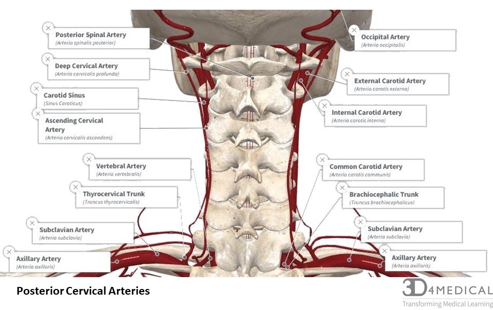
Common Carotid Arteries: The right common carotid arises from the brachiocephalic artery (brachio, BRĀ-key-ō, upper limb / cephalic, SĔF-ă-lik, pertaining to the head) while the left carotid artery branches directly from the aortic arch. Both bifurcate (split) into the internal and external arteries that supply blood to the face, skull, and brain.
Ascending Cervical Artery: The Ascending Cervical Artery runs anterior to transverse processes of the cervical vertebrae in a superior direction to provide blood supply to the muscles of the neck, vertebrae, and the vertebra canal.
Occipital Arteries: Send blood the to meninges, scalp and cervical muscles (mastoid and sternocleidomastoid).
VEINS:
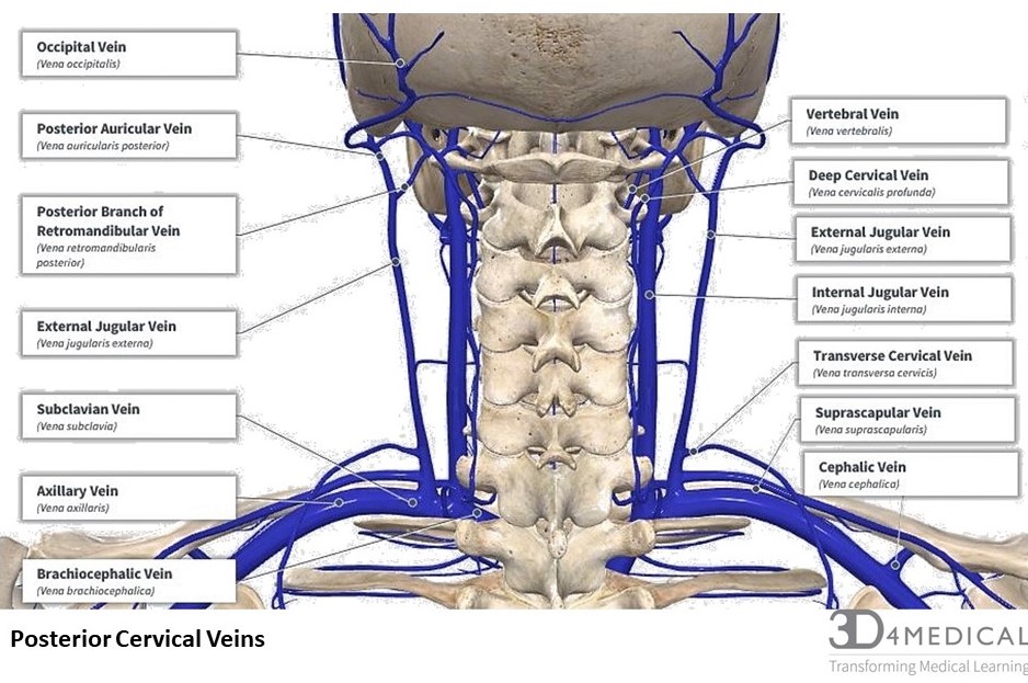
Internal Jugulars (right and left) are formed by the sigmoid and inferior petrosal venous sinuses and then descend to join with the brachiocephalic veins. The internal jugulars are the main venous return from the brain, face, and neck.
External Jugular Veins (right and left) are formed by posterior auricular and retromandibular veins and are joined by the transverse cervical vein, suprascapular and anterior jugular before merging with the subclavian vein.
Deep Cervical Veins are formed via anastomoses of the occipital veins and other small veins in the cervical region muscles such as the semispinalis capitis and the longissimus capitis. Before draining into the brachiocephalic vein, they join with the vertebral vein.
Occipital Veins (right and left) are superficial to the occipital fascia. Small vessels on the posterior of the scalp form the occipital veins before the descending to join the deep cervical, posterior auricular and parietal branch of the superficial temporal vein via anastomoses to form the external jugular. The occipital veins drain the muscles and skin of the posterior skull.
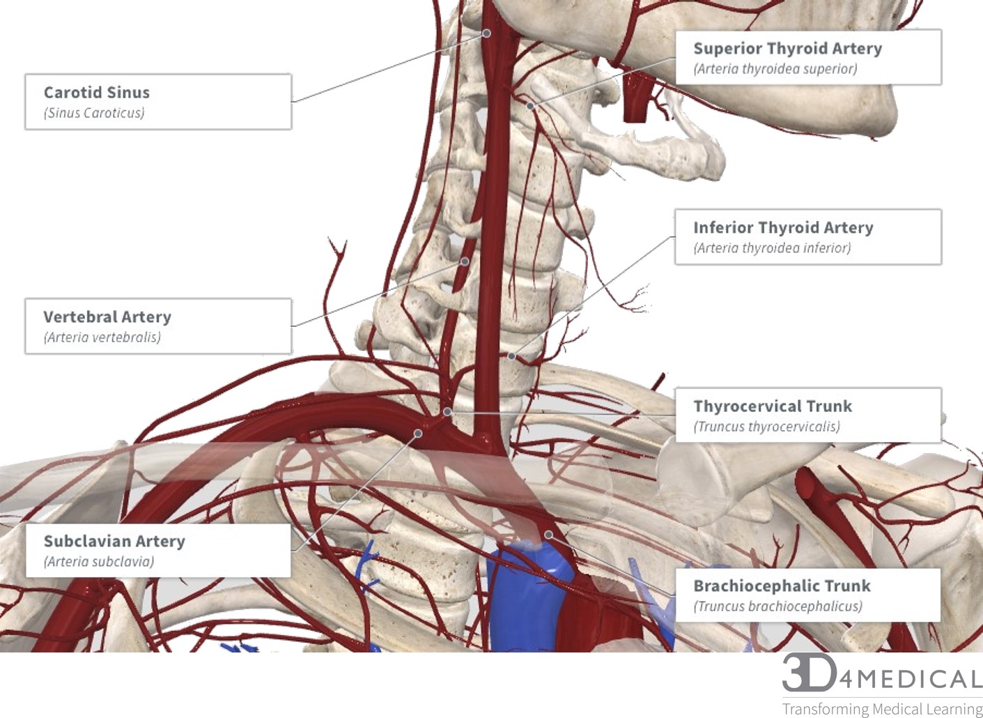
ANTERIOR CERVICAL ARTERIES
The arteries of the anterior neck surround the thyroid and other structures. They supply oxygenated blood to the laryngeal muscles as well as the supra and infrahyoid muscles. Starting from the aorta, the oxygenated blood will branch off to the brachiocephalic artery on the right side. This artery is unique as the left side does not have this feature. After the brachiocephalic artery, on the right side, the left and right sides divide into the common carotid, subclavian, and vertebral arteries. The subclavian artery goes to the arm and the vertebral artery goes to the brain along with the common carotid. For more information on what muscles are supplied by their respective artery, see the blood supply column in the muscles section of this region.
ANTERIOR CERVICAL VEINS
The veins of the anterior cervical region are very similar to the arteries as they are named because of their region. Starting with the superior vena cava, the vein divides into the right and left brachiocephalic veins. The brachiocephalic vein then divides into the subclavian, internal jugular, and external jugular.
LYMPH
In the posterior cervical region, the Superficial Cervical Lymph Vessels drain the superficial portions of the neck and pass inferiorly to the jugular trunk deep to Platysma in the posterior cervical triangle.
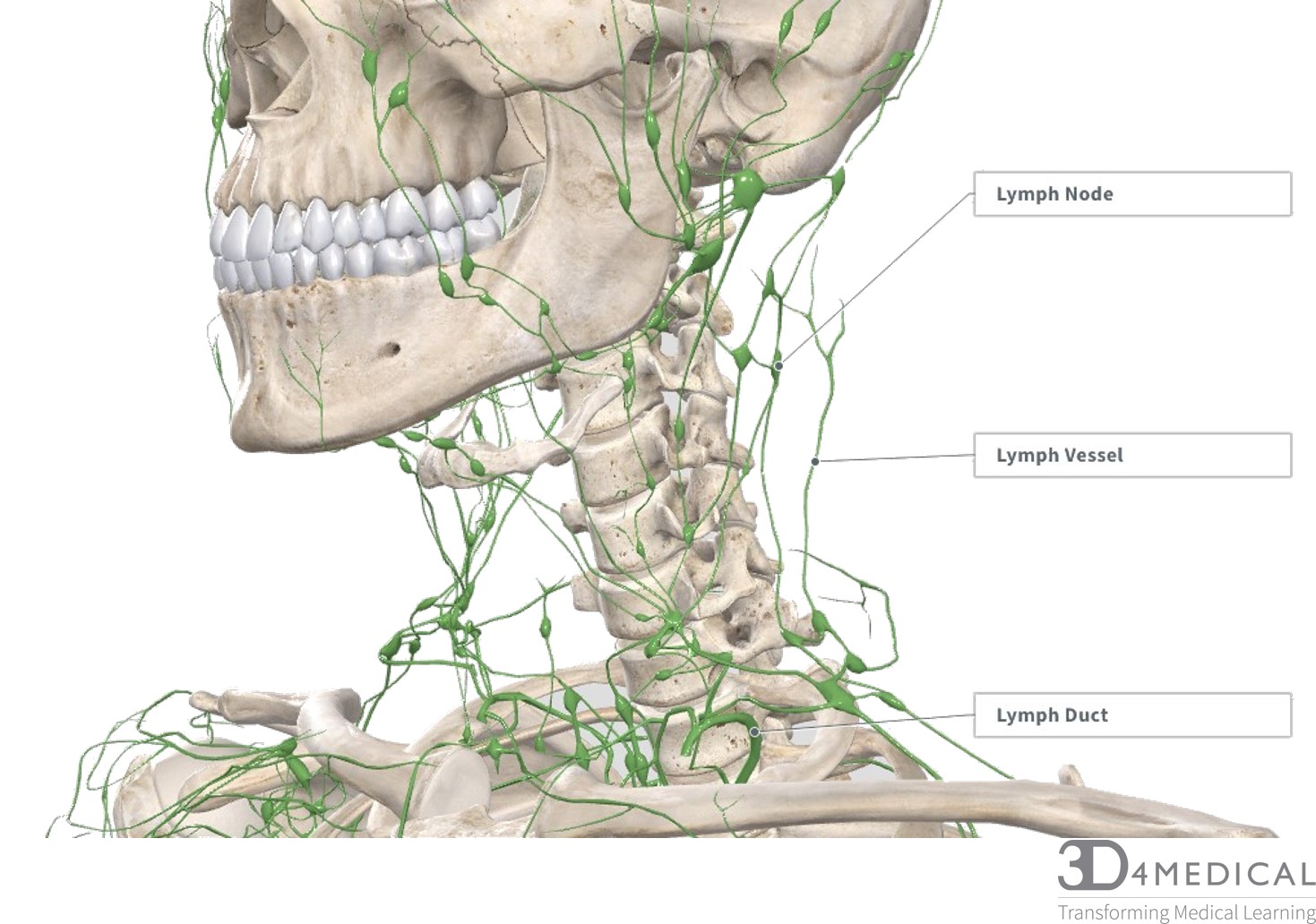
The lymphatic system is an essential part of the immune system. To simplify the system, it can be divided into 5 parts: vessels, valves, ducts, nodes, and organs. The picture below shows a vessels, duct, and node. There are many of these in the body and they are positioned uniquely to their region. Regardless of their position, they all have common purposes: to drain lymph to their respective organ(s), form plasma in the blood, and filter the lymph fluid.
Vertebral Artery:
Deep Cervical Artery:
