Introduction
Medical Imaging
X-ray
X-rays are the oldest and most robust modality of medical imaging. Developed and designed in 1895, X-rays have developed and allowed medical professions a broader understanding of various injuries resulting in bone damage, tumour growths and, infections to various tissues. X-rays are designed to produce images, which are constructed by electromagnetic waves called ionizing radiation. These waves or beam of radiation, pass through a determined part of the body. This in turn, allows the X-rays to be absorbed or scattered by internal structures; the remaining X-ray pattern is transmitted to a detector. Depending of the density of the structure, tissues will absorb different amounts of radiation depending on the regions density. Dense structures such as bone, will show up white on X-ray images; thinner structures or empty spaces, will appear black. Depending on the quality of the image, they may need to be enhanced in order to comprehend the various structures involved in the region. Solutions such as barium or iodine temporarily change how X-rays relate within the body. This results in causing certain structures to appear enhanced and highlighted. Compared to other imaging techniques, X-rays allow a diverse understanding of the structures within a region.

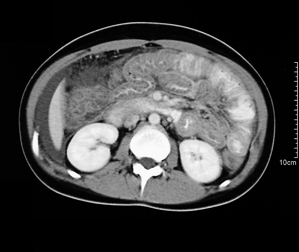
CT scan
Computerized Tomography or commonly known as CT scans, was introduced in 1971 and have evolved exponentially since. CT scans are programmed to integrate a sequence of X-ray images which are acquired from various angles that surround your body. Furthermore, CT scans uses computer processing programs to reassemble cross sectional images or slices. This medical imaging modality is very similar to X-ray imaging withholding the exception that countless X-ray beams and electronic X-ray detectors circulate around the patient, measuring the amount of radiation being absorbed by your body. The next step results in the computer program generating accurate and detailed two-dimensional images, based on the data that’s been accumulated; CT scans are also used to help detect a variety of diseases or conditions. Out of the various technological, medial advances, it’s said to be one of the fastest and most accurate modalities
MRI Scan
Magnetic Resonance Imaging or MRI, allows us to visualize nearly every internal system without invading the internal systems and structures. Developed and created by Raymond Damadian, MRI`s have changed various procedures associated with medical imaging. Current MRI technology now consists of powerful magnetic fields and radio frequency pulses which penetrate through the body, resulting in images of the various structures. Therefore, such images generate precise images and unlike CT and X-ray, MRI uses no ionizing radiation. This allows prolonged exposure time as more complex scans can last over an hour. Radio frequency pulses re-align hydrogen atoms that naturally exist within the body. As the hydrogen atoms return to their regular alignment they emit distinct amounts of energy depending on the type of tissue they are in. A MR scanner captures this energy and processes it to build a picture of the tissues scanned.
Ultrasound
Ultrasound is sound with a frequency above the limit of human hearing. Sonography (the medical use of ultrasound) uses these sound waves to produce images of the inside of the body. A transducer sends high frequency sound waves into the body then listens for the returning echoes to bounce back. When a sound wave strikes an object it bounces back, or echoes. The transducer measures these echoes by direction and time then creates a real time image, enabling the operator to see movement and structure. Doppler ultrasound is a special technique (used mostly in ultrasounds of the heart or echocardiograms) that allows the operator to see and evaluate blood flow through arteries and veins. Doppler measures the direction and speed of blood cells as they move through the vessel. This movement causes a change in the frequency of the reflected sound waves, this is called the Doppler effect. Sonography is noninvasive and doesn’t involve any ionizing radiation which makes it a popular modality to measure fetal development. A few drawbacks associated with sonography include its limitation to penetrate through bone, air/gas and large amounts of adipose tissue.

